-
Primary Antibodies
- Cancer Research
- Immunology
- Microbiology
-
Signal Transduction
- Phosphodiesterases
- Receptor Interacting Proteins
- Adenylate Cyclases
- G-Protein Coupled Receptors
- Guanylate Cyclases & GTPases
- Protein Phosphatases
- Second Messengers
- Growth Factors & Hormones
- Cell Adhesion
- Calcium Binding
- Regulation
- Protein Transport
- Plasma Membrane
- Cytoskeleton / ECM
- Adapters
- ... More
- Metabolism & Homeostasis
- Protein Kinases
- Vision & Olfaction
- Channels & Transporters
- Epigenetics
- Stem Cells
- Cell Biology
- Neuroscience
- Cardiology
- Endocrine System
- Kits & Reagents
- Lab Services
- About us
- Contact us
- Distributors
Home page Validation
Validation
|
Related
|
Reactivity, performance and maintaining up-to-date accurate data are some of the reasons we produce high quality products.
Our quality control processes are enforced strictly using a variety of applications including ELISA, Immunohistochemistry, and Western Blot.
For all of our antibodies, proteins peptides, reagents and kits we constantly are enforcing our quality controls to ensure our products remain to be high quality. We ensure our findings are visible through our website, datasheet and technical support.
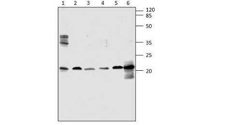
Western Blot of ARF4-401AP with various rat tissue samples and ARF4 positive control. 1) Rat hippocampus 2) Rat lung 3) PC-ARF4 4) Rat Spleen 5) Rat kidney. 1:250 dilution in DiluOBuffer.
Whether we are developing a custom antibody for you or if you simply ordered a trial size antibody, we will closely work with you to ensure you are getting the results you expect from your experiment. We happily publish, with permission, any validation data we receive through our collaborations to supplement our own findings.
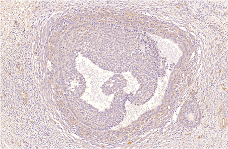
IHC of PD1B-201AP with human ovarian medulla tissue. 1:200 antibody dilution. Image courtesy of Steven S Petersen, MD, PhD Copenhagen University Hospital, Denmark.
Latest scientific knowledge
Our team of dedicated scientists research scientific papers to allow us to incorporate advances in our stringent quality controls. By comparing the results we obtain in our facility with recent scientific publications we ensure the data we post about our products is relevant and up to date.
We also actively collect publications using our products and publish the citations on our website. Oftentimes specialized testing conditions and results can be examined further in these publications
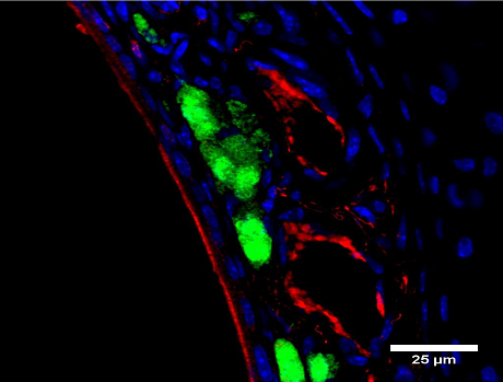
IF of PGCB-201AP with thin section of mouse nose tissue. Green: Grueneberg ganglion neurons. Red: antibody immunoreactivity. Blue: Nuclei. Staining is found on blood vessel walls. Data provided by Dr. Kroos lab; Caltech, Pasadena, CA.
|
Select your currency:
(c) FabGennix International Inc.