Your cart is empty.

Home page Primary Antibodies Immunology Innate Immunity Cytokines cKit Antibody
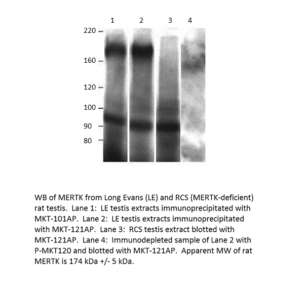
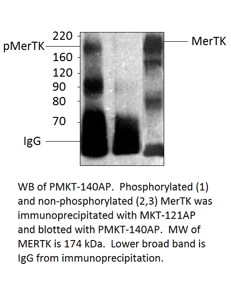
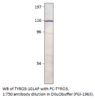
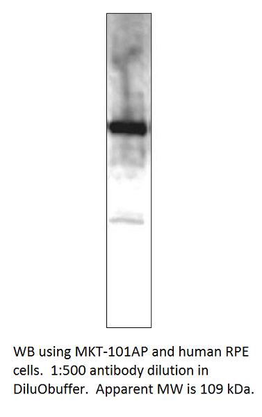
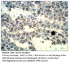
Home page Primary Antibodies Protein Kinases MERTK Antibody
Home page Primary Antibodies Protein Kinases MERTK Antibody
Home page Primary Antibodies Protein Kinases MERTK Antibody
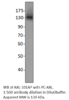
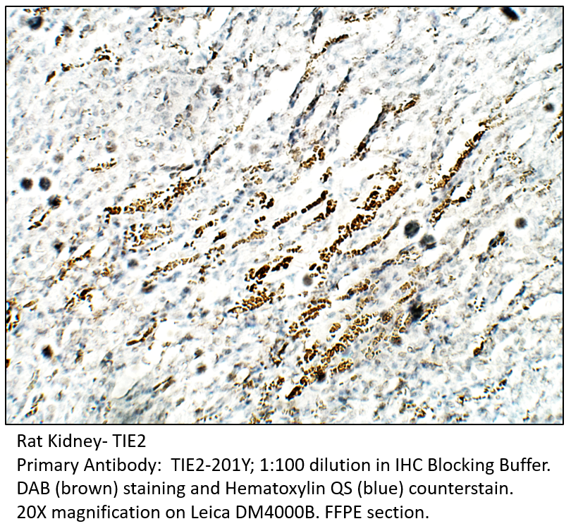
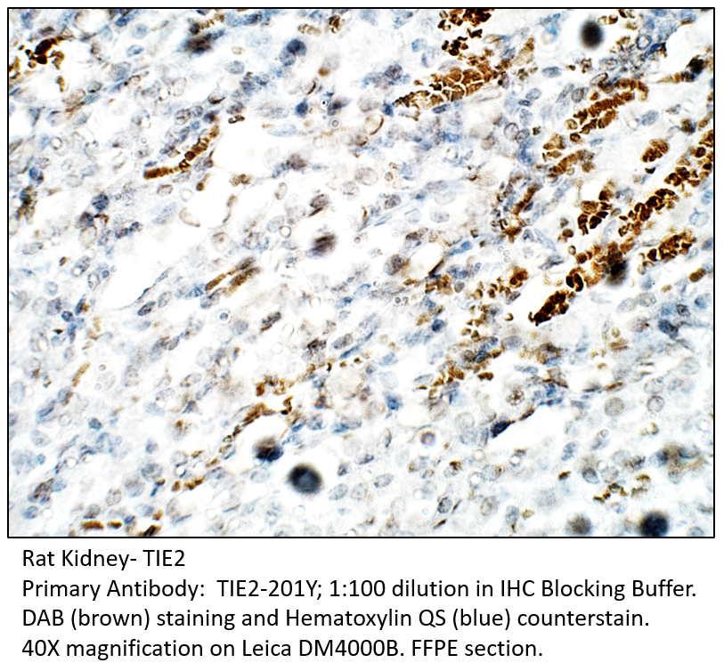
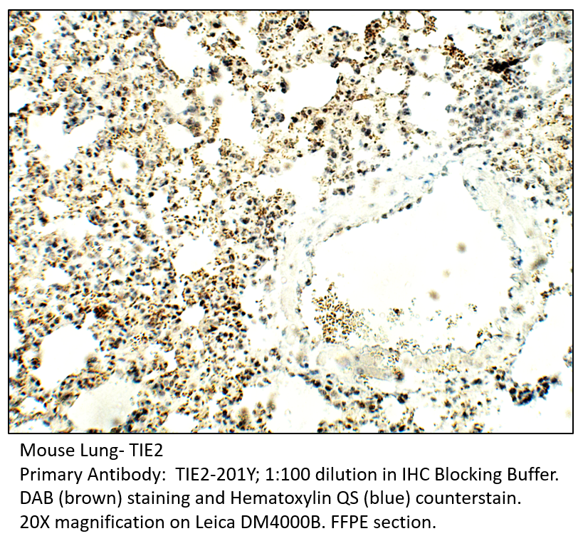
Home page Primary Antibodies Protein Kinases TIE2 Antibody
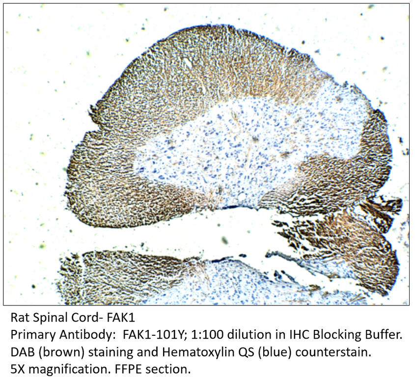
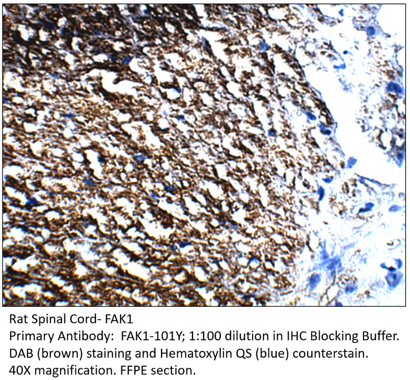
Home page Primary Antibodies Signal Transduction Cytoskeleton / ECM Integrins FAK1 Antibody
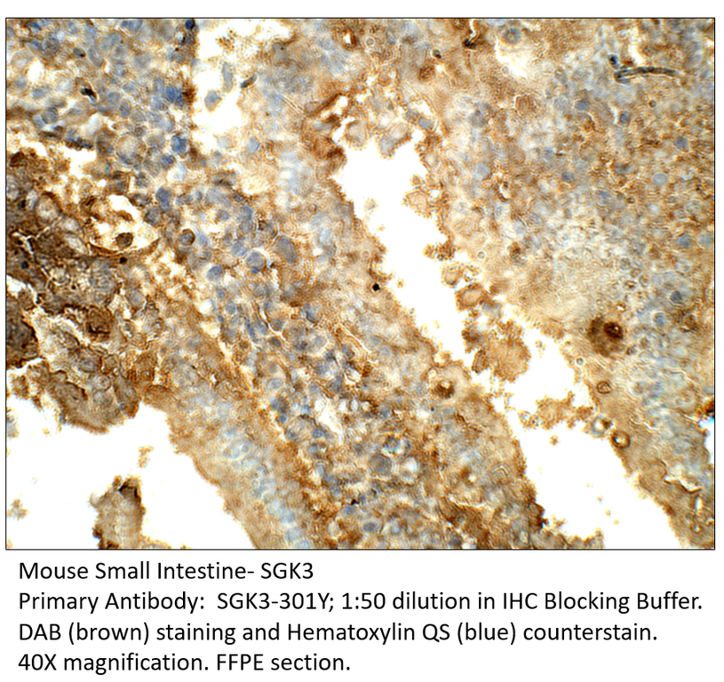
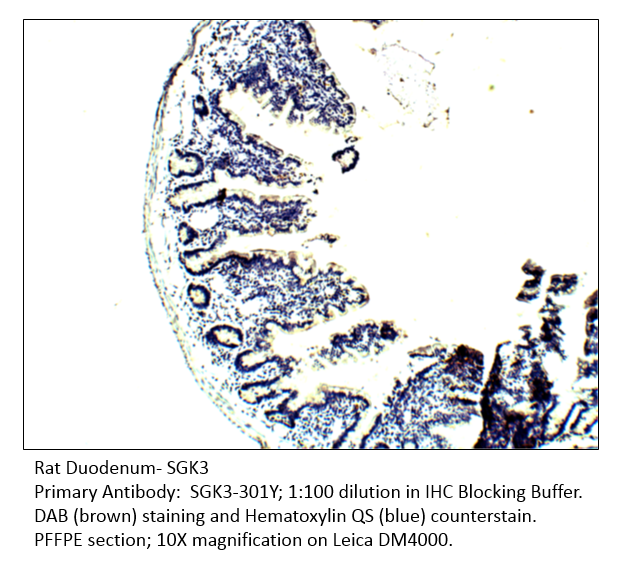
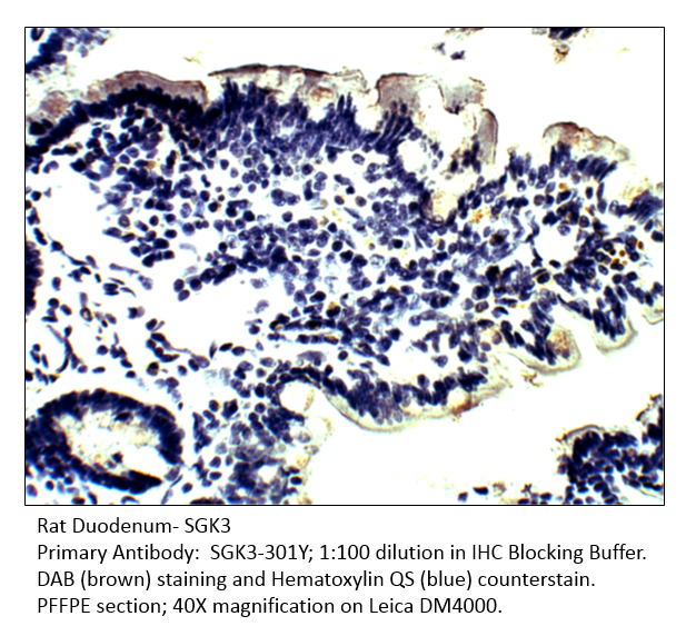
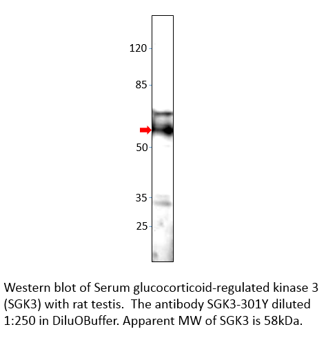
Home page Primary Antibodies Protein Kinases Serine/Threonine Kinases SGK3 Antibody
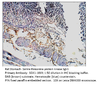
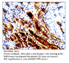
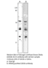
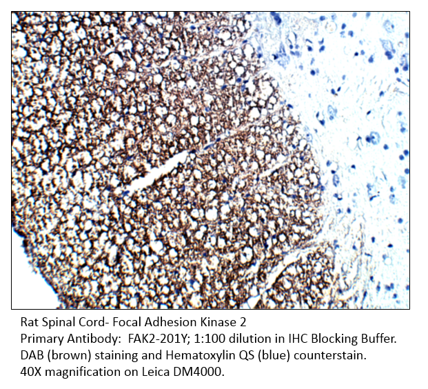
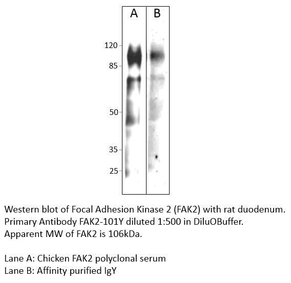
Home page Primary Antibodies Protein Kinases Tyrosine Kinases FAK2 Antibody
Home page Primary Antibodies Cardiology Angiogenesis Ephrins Ephrin Receptor A1 Antibody
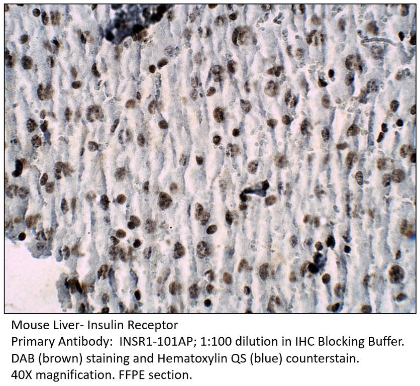
Home page Primary Antibodies Protein Kinases Tyrosine Kinases Insulin Receptor Antibody

Accessories
| Product | Note | Status | Price | |
|---|---|---|---|---|
|
CKIT.131-FITC | |||
|
CKIT.131-BIOTIN | |||
|
P-CKIT.131 | |||
|
PC-CKIT | |||
| Display accessory details | ||||
 cKit Antibody FITC
 cKit Antibody BIOTIN
 cKit Blocking Peptide
 cKit Positive Control
We also recommend
|
CKIT-141AP
 |
CKIT-101AP
 |
CKIT-112AP
 |
|
CKIT-121AP
 |
Browse these categories as well: Cytokines, Diagnostic Markers, Tumor Associated, Tyrosine Kinases, Channels & Transporters, Embryonic Germ Cells, Surface Molecules, Growth Factors, Growth Factors & Hormones, Oncoproteins, Mast Cell Lineage, Hematopoietic Progenitors, Reproduction, Organogenesis, Primaries, Receptor Tyrosine Kinases, Non-Receptor Tyrosine Kinases
Accessories
| Product | Note | Status | Price | |
|---|---|---|---|---|
|
MKT120-FITC | |||
|
MKT120-BIOTIN | |||
|
P-MKT120 | |||
|
PC-MKT | |||
| Display accessory details | ||||
 MERTK Antibody FITC
 MERTK Antibody BIOTIN
 MERTK Blocking Peptide
 MERTK Positive Control
We also recommend
|
MKT-112AP
 |
MKT-101AP
 |
PMKT-140AP
 |
|
TYRO3-101AP
 |
Customers who bought this product also bought
|
|
Browse these categories as well: Protein Kinases, Tyrosine Kinases, Vision, Vision & Olfaction, Neurotransmission, Primaries, Receptor Tyrosine Kinases

Accessories
| Product | Note | Status | Price | |
|---|---|---|---|---|
|
MKT110-FITC | |||
|
MKT110-BIOTIN | |||
|
P-MKT110 | |||
|
PC-MKT | |||
| Display accessory details | ||||
 MERTK Antibody FITC
 MERTK Antibody BIOTIN
 MERTK Blocking Peptide
We also recommend
|
MKT-101AP
|
MKT-121AP
 |
PMKT-140AP
|
|
TYRO3-101AP
|
Browse these categories as well: Protein Kinases, Tyrosine Kinases, Vision & Olfaction, Vision, Neurotransmission, Primaries, Receptor Tyrosine Kinases

Accessories
| Product | Note | Status | Price | |
|---|---|---|---|---|
|
MKT100-FITC | |||
|
MKT100-BIOTIN | |||
|
P-MKT100 | |||
|
PC-MKT | |||
| Display accessory details | ||||
 MERTK Antibody FITC
 MERTK Antibody BIOTIN
 MERTK Blocking Peptide
We also recommend
|
MKT-112AP
|
MKT-121AP
|
PMKT-140AP
|
|
AXL-101AP
 |
TYRO3-101AP
|
Browse these categories as well: Protein Kinases, Tyrosine Kinases, Vision, Vision & Olfaction, Neurotransmission, Primaries, Receptor Tyrosine Kinases


Accessories
| Product | Note | Status | Price | |
|---|---|---|---|---|
|
TIE2-Y-FITC | |||
|
TIE2-Y-BIOTIN | |||
|
P-TIE2-Y | |||
|
PC-TIE2 | |||
| Display accessory details | ||||
 TIE2 Antibody FITC
 TIE2 Antibody BIOTIN
 TIE2 Blocking Peptide
 TIE2 Positive Control
We also recommend
|
Anti-Chicken Secondary
 |
Rabbit Polyclonal
 |
Browse these categories as well: Protein Kinases, Tyrosine Kinases, Cardiology, Oncoproteins, Growth Factors, Growth Factors & Hormones, Organogenesis, Cardiovascular Markers, Primaries, Chicken IgY Primaries
Accessories
| Product | Note | Status | Price | |
|---|---|---|---|---|
|
FAK1-Y-FITC | |||
|
FAK1-Y-BIOTIN | |||
|
P-FAK1-Y | |||
|
PC-FAK1 | |||
| Display accessory details | ||||
 FAK1 Antibody FITC
 FAK1 Antibody BIOTIN
 FAK1 Blocking Peptide
 FAK1 Positive Control
We also recommend
|
Anti-Chicken Secondary
|
FAK2-201Y
 |
Browse these categories as well: Integrins, Tyrosine Kinases, Cell Adhesion, Signal Transduction, Protein Kinases, Primaries, Chicken IgY Primaries, Non-Receptor Tyrosine Kinases
Accessories
| Product | Note | Status | Price | |
|---|---|---|---|---|
|
SGK3-Y-FITC | |||
|
SGK3-Y-BIOTIN | |||
|
P-SGK3-Y | |||
|
PC-SGK3 | |||
| Display accessory details | ||||
 SGK3 Antibody FITC
 SGK3 Antibody BIOTIN
 SGK3 Blocking Peptide
 SGK3 Positive Control
We also recommend
|
Anti-Chicken Secondary
|
SGK1-101Y
 |
SGK2-201Y
 |
|
GSK3B-301Y
 |
Browse these categories as well: Serine/Threonine Kinases, Tyrosine Kinases, Protein Kinases, Amino Acid Metabolism, Chicken IgY Primaries, Primaries
Accessories
| Product | Note | Status | Price | |
|---|---|---|---|---|
|
FAK2-Y-FITC | |||
|
FAK2-Y-BIOTIN | |||
|
P-FAK2-Y | |||
|
PC-FAK2 | |||
| Display accessory details | ||||
 FAK2 Antibody FITC
 FAK2 Antibody BIOTIN
 FAK2 Blocking Peptide
 FAK2 Positive Control
We also recommend
|
Anti-Chicken Secondary
|
FAK1-101Y
 |
Browse these categories as well: Tyrosine Kinases, Hypoxia, Metabolism, Primaries, Chicken IgY Primaries, Non-Receptor Tyrosine Kinases

Accessories
| Product | Note | Status | Price | |
|---|---|---|---|---|
|
EPHAR1-FITC | |||
|
EPHAR1-BIOTIN | |||
|
P-EPHAR1 | |||
|
PC-EPHAR1 | |||
| Display accessory details | ||||
 Ephrin Receptor A1 Antibody FITC
 Ephrin Receptor A1 Antibody BIOTIN
 Ephrin Receptor A1 Blocking Peptide
 Ephrin Receptor A1 Positive Control
We also recommend
|
EPHBR1-101AP
 |
Browse these categories as well: Ephrins, Protein Kinases, Tyrosine Kinases, Cell markers & CAMs, Neurogenesis & Plasticity, Neurology process, Neurotransmission, Channels & Transporters, Primaries, Receptor Tyrosine Kinases
Accessories
| Product | Note | Status | Price | |
|---|---|---|---|---|
|
INSR1-FITC | |||
|
INSR1-BIOTIN | |||
|
P-INSR1 | |||
|
PC-INSR1 | |||
| Display accessory details | ||||
 Insulin Receptor Antibody FITC
 Insulin Receptor Antibody BIOTIN
 Insulin Receptor Blocking Peptide
 Insulin Receptor Positive Control
We also recommend
|
INSRB-201AP
 |
INSR1-112AP
 |
Browse these categories as well: Tyrosine Kinases, Insulin, Metabolism, Metabolism & Homeostasis, Growth Factors, Growth Factors & Hormones, Neurology process, Atherosclerosis, Diabetes, Primaries