Your cart is empty.

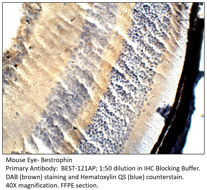
Home page Primary Antibodies Neuroscience Bestrophin Antibody
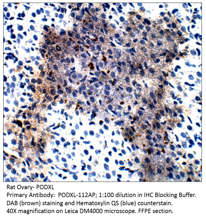
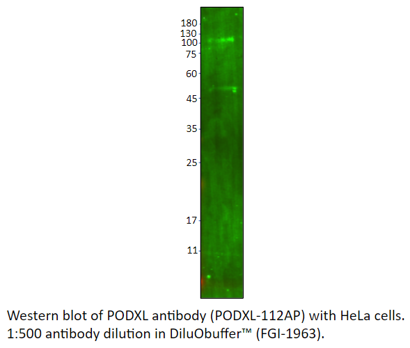
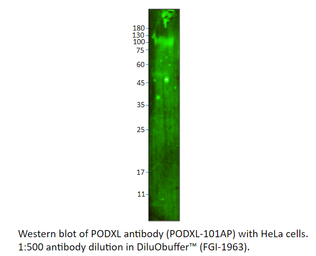
Home page Primary Antibodies Signal Transduction Cell Adhesion PODXL Antibody
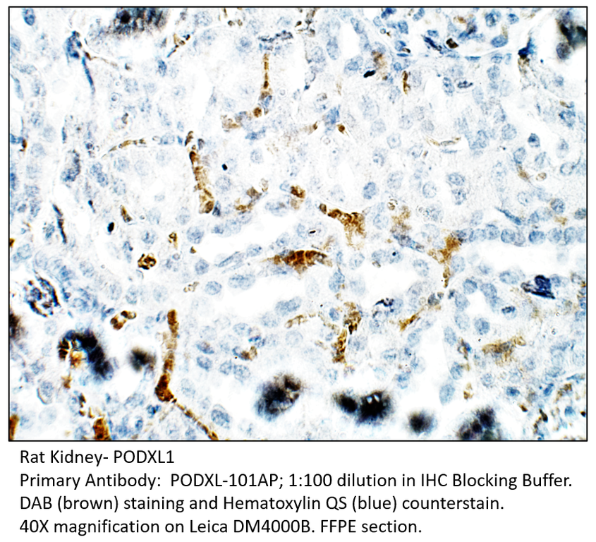
Home page Primary Antibodies Signal Transduction Cell Adhesion PODXL1 Antibody
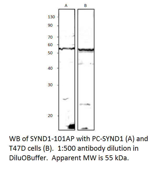
Home page Primary Antibodies Signal Transduction Cell Adhesion Syndecan-1 Antibody
Home page Primary Antibodies Signal Transduction Cell Adhesion Syndecan-1 Antibody
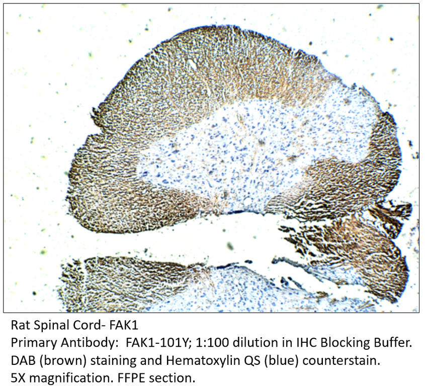
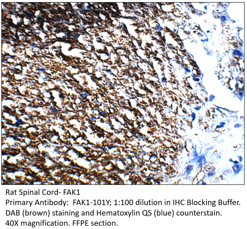
Home page Primary Antibodies Signal Transduction Cytoskeleton / ECM Integrins FAK1 Antibody
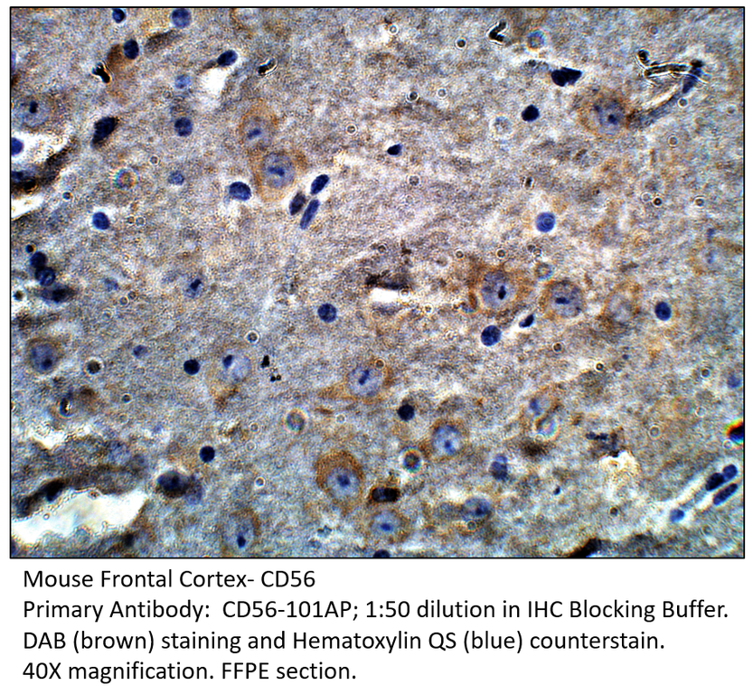
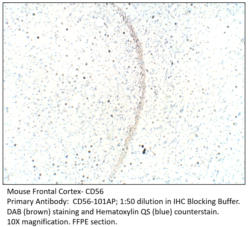
Home page Primary Antibodies Neuroscience Cell markers & CAMs CD56 / NCAM Antibody
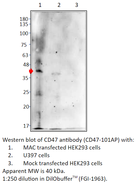
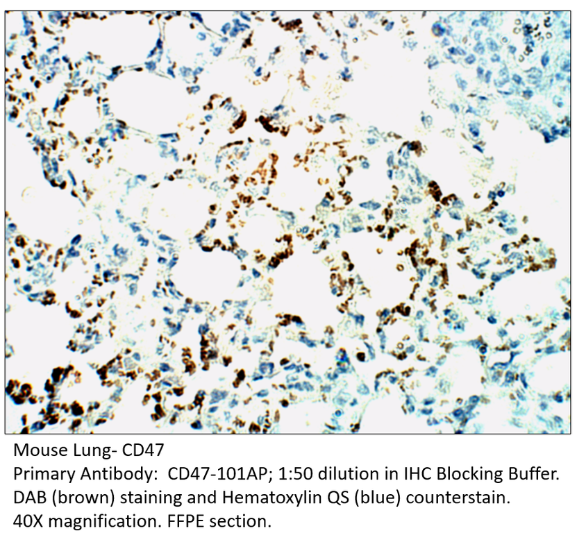
Home page Primary Antibodies Signal Transduction Cell Adhesion CD47 Antibody
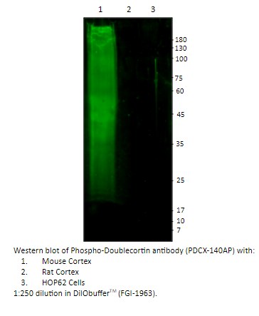
Home page Primary Antibodies Signal Transduction Cytoskeleton / ECM Microtubules Phospho-Doublecortin Antibody
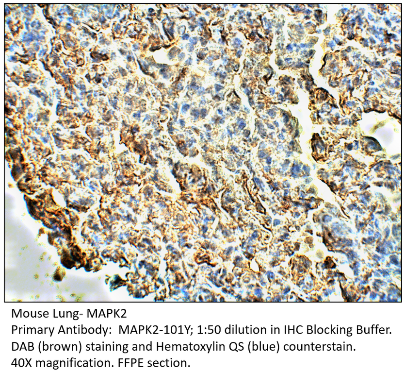
Home page Primary Antibodies Neuroscience Neurodegenerative Disease Alzheimer's Research MAPK2 Antibody
Accessories
| Product | Note | Status | Price | |
|---|---|---|---|---|
|
BEST1.3-FITC | |||
|
BEST1.3-BIOTIN | |||
|
P-BEST1.3 | |||
|
PC-BEST1 | |||
| Display accessory details | ||||
 Bestrophin Antibody FITC
 Bestrophin Antibody BIOTIN
 Bestrophin Blocking Peptide
 Bestrophin Positive Control
We also recommend
|
BEST-101AP
 |
BEST-112AP
 |
BEST-301AP
 |
|
BEST-312AP
 |
Browse these categories as well: Neuroscience, ECM Proteins, Cell Adhesion, Vision, Vision & Olfaction, Primaries

Accessories
| Product | Note | Status | Price | |
|---|---|---|---|---|
|
PODXL.112-FITC | |||
|
PODXL.112-BIOTIN | |||
|
P-PODXL.112 | |||
|
PC-PODXL | |||
| Display accessory details | ||||
 PODXL Antibody FITC
 PODXL Antibody BIOTIN
 PODXL Blocking Peptide
 PODXL Positive Control
We also recommend
|
PODXL-101AP
 |
Browse these categories as well: Cell Adhesion, Diagnostic Markers, Extra Cellular Matrix, Surface Molecules, Cardiology, Primaries
Accessories
| Product | Note | Status | Price | |
|---|---|---|---|---|
|
PODXL.101-FITC | |||
|
PODXL.101-BIOTIN | |||
|
P-PODXL.101 | |||
|
PC-PODXL | |||
| Display accessory details | ||||
 PODXL1 Antibody FITC
 PODXL1 Antibody BIOTIN
 PODXL1 Blocking Peptide
We also recommend
|
PODXL-112AP
 |
Browse these categories as well: Cell Adhesion, Diagnostic Markers, Extra Cellular Matrix, Surface Molecules, Cardiology, Primaries

Accessories
| Product | Note | Status | Price | |
|---|---|---|---|---|
|
SYND1.112-FITC | |||
|
SYND1.112-BIOTIN | |||
|
P-SYND1.112 | |||
|
PC-SYND1 | |||
| Display accessory details | ||||
 Syndecan-1 Antibody FITC
 Syndecan-1 Antibody BIOTIN
 Syndecan-1 Blocking Peptide
 Syndecan-1 Positive Control
We also recommend
|
SYND1-101AP
 |
Browse these categories as well: Cell Adhesion, Diagnostic Markers, ECM Proteins, Cell markers & CAMs, Neurogenesis & Plasticity, Invasion/microenviornment, B Lymphocytic Lineage, Lipids, Metabolism & Homeostasis, Metabolism, Primaries
Accessories
| Product | Note | Status | Price | |
|---|---|---|---|---|
|
SYND1.101-FITC | |||
|
SYND1.101-BIOTIN | |||
|
P-SYND1.101 | |||
|
PC-SYND1 | |||
| Display accessory details | ||||
 Syndecan-1 Antibody FITC
 Syndecan-1 Antibody BIOTIN
 Syndecan-1 Blocking Peptide
We also recommend
|
SYND1-112AP
 |
Browse these categories as well: Cell Adhesion, Diagnostic Markers, ECM Proteins, Cell markers & CAMs, Neurogenesis & Plasticity, Invasion/microenviornment, B Lymphocytic Lineage, Lipids, Metabolism & Homeostasis, Metabolism, Primaries
Accessories
| Product | Note | Status | Price | |
|---|---|---|---|---|
|
FAK1-Y-FITC | |||
|
FAK1-Y-BIOTIN | |||
|
P-FAK1-Y | |||
|
PC-FAK1 | |||
| Display accessory details | ||||
 FAK1 Antibody FITC
 FAK1 Antibody BIOTIN
 FAK1 Blocking Peptide
 FAK1 Positive Control
We also recommend
|
Anti-Chicken Secondary
 |
FAK2-201Y
 |
Browse these categories as well: Integrins, Tyrosine Kinases, Cell Adhesion, Signal Transduction, Protein Kinases, Primaries, Chicken IgY Primaries, Non-Receptor Tyrosine Kinases


Accessories
| Product | Note | Status | Price | |
|---|---|---|---|---|
|
CD56-FITC | |||
|
CD56-BIOTIN | |||
|
P-CD56 | |||
|
PC-CD56 | |||
| Display accessory details | ||||
 CD56 Antibody FITC
 CD56 Antibody BIOTIN
 CD56 Blocking Peptide
 CD56 Positive Control
Browse these categories as well: Cell markers & CAMs, Diagnostic Markers, ECM Proteins, Cell Adhesion, Neurology process, Hematopoietic Progenitors, Primaries, CD Markers
Accessories
| Product | Note | Status | Price | |
|---|---|---|---|---|
|
CD47-BIOTIN | |||
|
CD47-FITC | |||
|
P-CD47 | |||
|
PC-CD47 | |||
| Display accessory details | ||||
 CD47 Antibody BIOTIN
 CD47 Antibody FITC
 CD47 Blocking Peptide
 CD47 Positive Control
Browse these categories as well: Cell Adhesion, CD Markers, Primaries
Accessories
| Product | Note | Status | Price | |
|---|---|---|---|---|
|
PDCX-BIOTIN | |||
|
PDCX-FITC | |||
|
P-PDCX | |||
|
PC-PDCX | |||
| Display accessory details | ||||
 Phospho-DoublecortinAntibody BIOTIN
 Phospho-Doublecortin Antibody FITC
 Phospho-Doublecortin Blocking Peptide
 Phospho-Doublecortin Positive Control
Browse these categories as well: Microtubules, Cell Adhesion, Tags and Cell Markers, Cell markers & CAMs, Primaries
Accessories
| Product | Note | Status | Price | |
|---|---|---|---|---|
|
MAPK2-BIOTIN | |||
|
MAPK2-FITC | |||
|
P-MAPK2 | |||
|
PC-MAPK2 | |||
| Display accessory details | ||||
 MAPK2 Antibody BIOTIN
 MAPK2 Antibody FITC
 MAPK2 Blocking Peptide
 MAPK2 Positive Control
We also recommend
|
Anti-Chicken Secondary
|
Browse these categories as well: Alzheimer's Research, MAPK Pathway, TGF Pathway, TGF beta, Cell Adhesion, Cytoskeleton / ECM, Apoptosis, Apoptosis, Primaries, Chicken IgY Primaries