|
General Information
|
|
|
Product
|
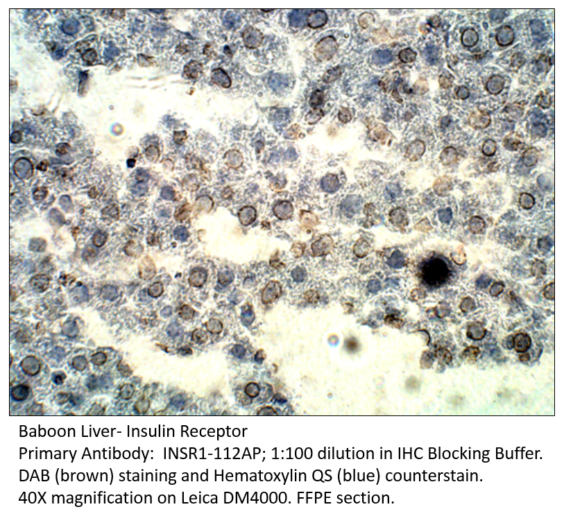
Insulin Receptor beta Antibody
|
|
Verified Applications
|
ELISA, IP, WB
|
|
Host
|
Rabbit
|
|
Species Cross Reactivity
|
Human, Mouse, Rat
|
|
Immunogen
|
Synthetic peptide taken within amino acid region 725-775 on human Insulin receptor beta subunit protein.
|
|
Physical Properties
|
|
|
Quantity
|
100 µg
|
|
Volume
|
200 µl
|
|
Concentration
|
0.55-0.75 µg/µl in antibody stabilization buffer
|
|
Immunoglobulin
|
IgG
|
|
Form
|
Affinity Purified
|
|
Clonality
|
Polyclonal
|
|
Storage
|
-20⁰C for long term storage
|
|
Application Protocol
|
|
ELISA
|
1:10,000
|
|
Immunoprecipitation
|
1:200
|
|
Western Blot
|
1:500
|
|
Protein
|
|
Uniprot #
|
P06213
|
|
Overview
|
Receptor tyrosine kinase which mediates the pleiotropic actions of insulin. Binding of insulin leads to phosphorylation of several intracellular substrates, including, insulin receptor substrates (IRS1, 2, 3, 4), SHC, GAB1, CBL and other signaling intermediates. Each of these phosphorylated proteins serve as docking proteins for other signaling proteins that contain Src-homology-2 domains (SH2 domain) that specifically recognize different phosphotyrosines residues, including the p85 regulatory subunit of PI3K and SHP2. Phosphorylation of IRSs proteins lead to the activation of two main signaling pathways: the PI3K-AKT/PKB pathway, which is responsible for most of the metabolic actions of insulin, and the Ras-MAPK pathway, which regulates expression of some genes and cooperates with the PI3K pathway to control cell growth and differentiation. Binding of the SH2 domains of PI3K to phosphotyrosines on IRS1 leads to the activation of PI3K and the generation of phosphatidylinositol-(3, 4, 5)-triphosphate (PIP3), a lipid second messenger, which activates several PIP3-dependent serine/threonine kinases, such as PDPK1 and subsequently AKT/PKB. The net effect of this pathway is to produce a translocation of the glucose transporter SLC2A4/GLUT4 from cytoplasmic vesicles to the cell membrane to facilitate glucose transport. Moreover, upon insulin stimulation, activated AKT/PKB is responsible for: anti-apoptotic effect of insulin by inducing phosphorylation of BAD; regulates the expression of gluconeogenic and lipogenic enzymes by controlling the activity of the winged helix or forkhead (FOX) class of transcription factors. Another pathway regulated by PI3K-AKT/PKB activation is mTORC1 signaling pathway which regulates cell growth and metabolism and integrates signals from insulin. AKT mediates insulin-stimulated protein synthesis by phosphorylating TSC2 thereby activating mTORC1 pathway. The Ras/RAF/MAP2K/MAPK pathway is mainly involved in mediating cell growth, survival and cellular differentiation of insulin. Phosphorylated IRS1 recruits GRB2/SOS complex, which triggers the activation of the Ras/RAF/MAP2K/MAPK pathway. In addition to binding insulin, the insulin receptor can bind insulin-like growth factors (IGFI and IGFII). Isoform Short has a higher affinity for IGFII binding. When present in a hybrid receptor with IGF1R, binds IGF1. PubMed:12138094 shows that hybrid receptors composed of IGF1R and INSR isoform Long are activated with a high affinity by IGF1, with low affinity by IGF2 and not significantly activated by insulin, and that hybrid receptors composed of IGF1R and INSR isoform Short are activated by IGF1, IGF2 and insulin. In contrast, PubMed:16831875 shows that hybrid receptors composed of IGF1R and INSR isoform Long and hybrid receptors composed of IGF1R and INSR isoform Short have similar binding characteristics, both bind IGF1 and have a low affinity for insulin.
|
|
Molecular Function
|
Kinase, Receptor, Transferase, Tyrosine-protein kinase
|
|
Subcellular Location
|
Cell membrane; Single-pass type I membrane protein
|
|
Expression
|
Isoform Long and isoform Short are predominantly expressed in tissue targets of insulin metabolic effects: liver, adipose tissue and skeletal muscle but are also expressed in the peripheral nerve, kidney, pulmonary alveoli, pancreatic acini, placenta vascular endothelium, fibroblasts, monocytes, granulocytes, erythrocytes and skin. Isoform Short is preferentially expressed in fetal cells such as fetal fibroblasts, muscle, liver and kidney. Found as a hybrid receptor with IGF1R in muscle, heart, kidney, adipose tissue, skeletal muscle, hepatoma, fibroblasts, spleen and placenta (at protein level). Overexpressed in several tumors, including breast, colon, lung, ovary, and thyroid carcinomas.
|
|
Structure
|
Tetramer of 2 alpha and 2 beta chains linked by disulfide bonds. The alpha chains carry the insulin-binding regions, while the beta chains carry the kinase domain. Forms a hybrid receptor with IGF1R, the hybrid is a tetramer consisting of 1 alpha chain and 1 beta chain of INSR and 1 alpha chain and 1 beta chain of IGF1R. Interacts with SORBS1 but dissociates from it following insulin stimulation. Binds SH2B2. Activated form of INSR interacts (via Tyr-999) with the PTB/PID domains of IRS1 and SHC1. The sequences surrounding the phosphorylated NPXY motif contribute differentially to either IRS1 or SHC1 recognition. Interacts (via tyrosines in the C-terminus) with IRS2 (via PTB domain and 591-786 AA); the 591-786 would be the primary anchor of IRS2 to INSR while the PTB domain would have a stabilizing action on the interaction with INSR. Interacts with the SH2 domains of the 85 kDa regulatory subunit of PI3K (PIK3R1) in vitro, when autophosphorylated on tyrosine residues. Interacts with SOCS7. Interacts (via the phosphorylated Tyr-999), with SOCS3. Interacts (via the phosphorylated Tyr-1185, Tyr-1189, Tyr-1190) with SOCS1. Interacts with CAV2 (tyrosine-phosphorylated form); the interaction is increased with 'Tyr-27'phosphorylation of CAV2 (By similarity). Interacts with ARRB2 (By similarity). Interacts with GRB10; this interaction blocks the association between IRS1/IRS2 and INSR, significantly reduces insulin-stimulated tyrosine phosphorylation of IRS1 and IRS2 and thus decreases insulin signaling. Interacts with GRB7. Interacts with PDPK1. Interacts (via Tyr-1190) with GRB14 (via BPS domain); this interaction protects the tyrosines in the activation loop from dephosphorylation, but promotes dephosphorylation of Tyr-999, this results in decreased interaction with, and phosphorylation of, IRS1. Interacts (via subunit alpha) with ENPP1 (via 485-599 AA); this interaction blocks autophosphorylation. Interacts with PTPRE; this interaction is dependent of Tyr-1185, Tyr-1189 and Tyr-1190 of the INSR. Interacts with STAT5B (via SH2 domain). Interacts with PTPRF. Interacts with ATIC; ATIC together with PRKAA2/AMPK2 and HACD3/PTPLAD1 is proposed to be part of a signaling netwok regulating INSR autophosphorylation and endocytosis (By similarity).
|
|
Alternative Nomenclature
|
CD 220 antibody
CD220 antigen antibody
HHF5 antibody
HIR B antibody
INSR antibody
Insulin receptor antibody
Insulin receptor subunit beta antibody
IR antibody
|
| |
|

 Insulin Receptor Antibody FITC
 Insulin Receptor Antibody BIOTIN
 Insulin Receptor Blocking Peptide
 Insulin Receptor Positive Control

 Insulin Receptor beta Antibody FITC
 Insulin Receptor beta Antibody BIOTIN
 Insulin Receptor beta Blocking Peptide
 Insulin Receptor beta Positive Control
 VEGFR1 Antibody FITC
 VEGFR1 Antibody BIOTIN
 VEGFR1 Blocking Peptide
 VEGFR1 Positive Control
 TrkA Antibody FITC
 TrkA Antibody BIOTIN
 TrkA Blocking Peptide
 TrkA Positive Control
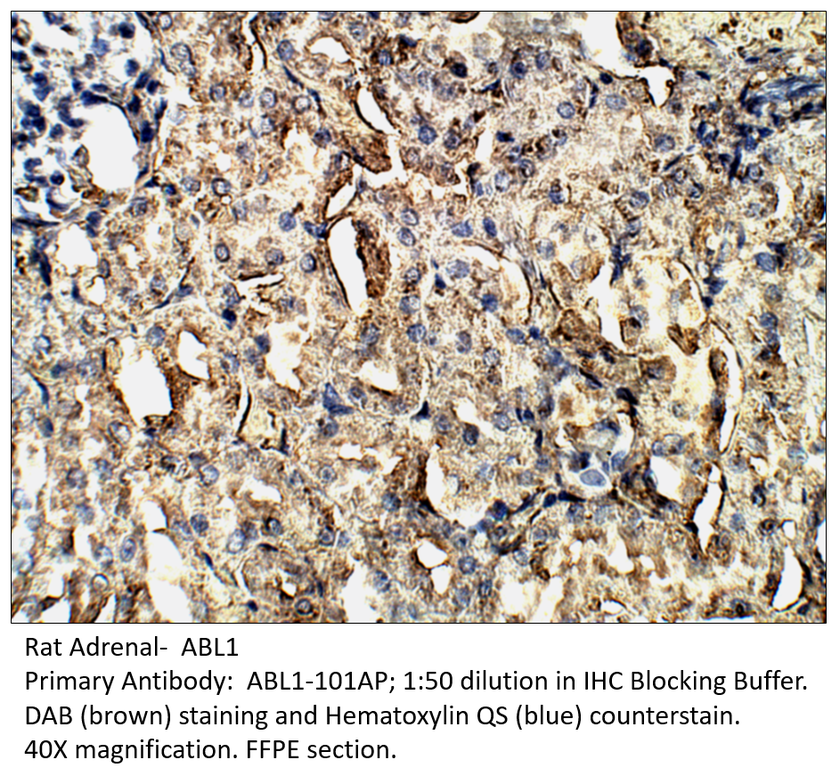
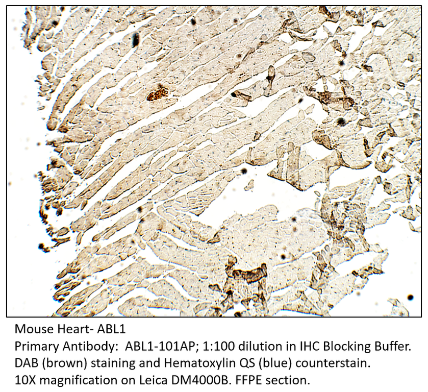
 ABL1 Antibody BIOTIN
 ABL1 Antibody FITC
 ABL1 Blocking Peptide
 ABL1 Positive Control
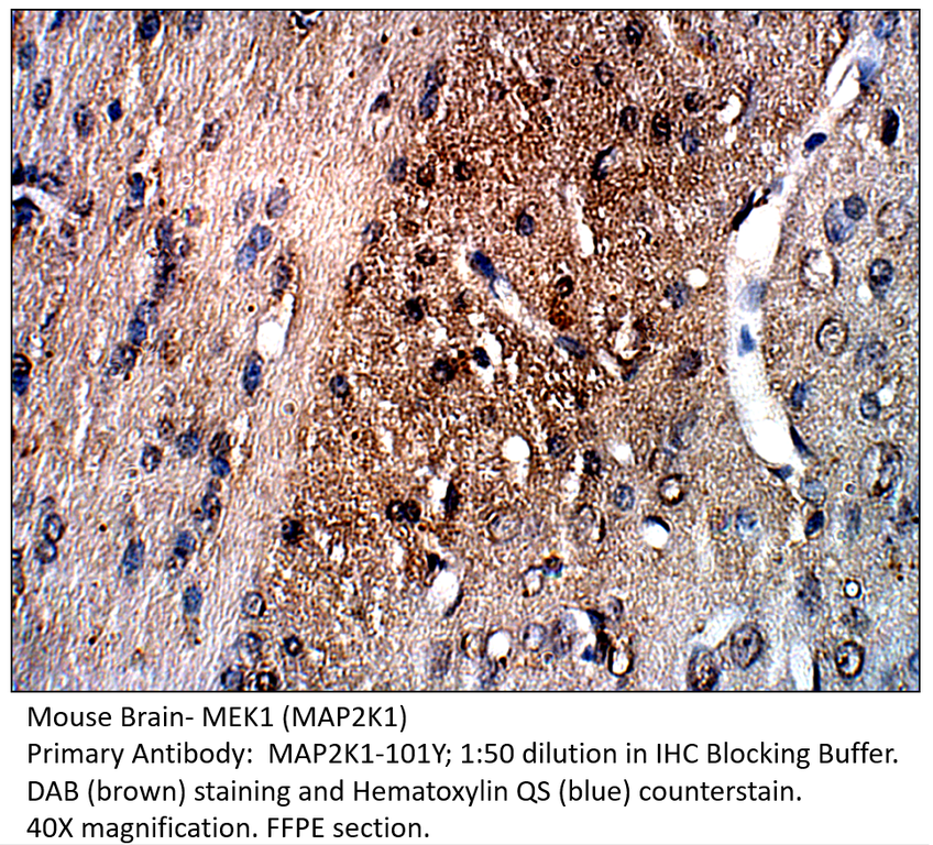
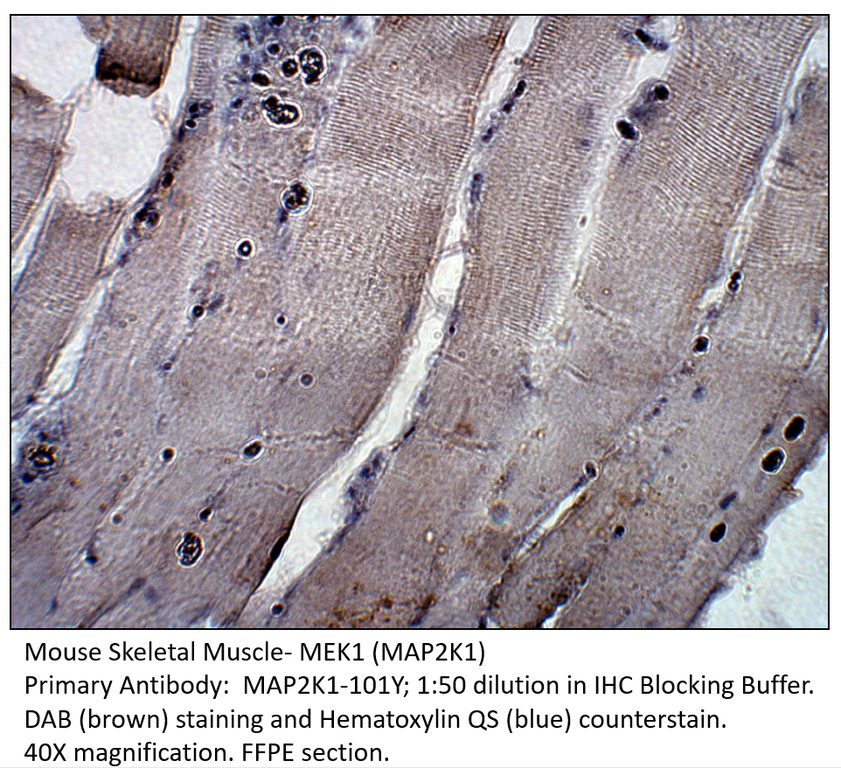
 MAP2K1 Antibody BIOTIN
 MAP2K1 Antibody FITC
 MAP2K1 Blocking Peptide
 Phospho-JAK1 Antibody BIOTIN
 Phospho-JAK1 Antibody FITC
 Phospho-JAK1 Blocking Peptide
 Phospho-JAK1 Positive Control
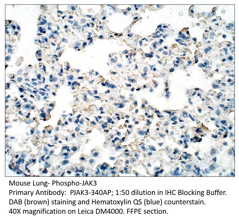
 Phospho-JAK3 Antibody BIOTIN
 Phospho-JAK3 Antibody FITC
 Phospho-JAK3 Blocking Peptide
 Phospho-JAK3 Positive Control

 LIMK1 Antibody BIOTIN
 LIMK1 Antibody FITC
 LIMK1 Blocking Peptide
 LIMK1 Positive Control

 TYK2 Antibody BIOTIN
 TYK2 Positive Control
 TYK2 Blocking Peptide
 TYK2 Antibody FITC