Your cart is empty.

Home page Primary Antibodies Protein Kinases Serine/Threonine Kinases PKC Phospho-PKC eta Antibody
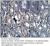
Home page Primary Antibodies Protein Kinases Serine/Threonine Kinases PKC Phospho-PKC eta Antibody
Home page Primary Antibodies Protein Kinases Serine/Threonine Kinases PKC PKC eta Antibody
Home page Primary Antibodies Protein Kinases Serine/Threonine Kinases PKC PKC eta Antibody
Home page Primary Antibodies Protein Kinases Serine/Threonine Kinases PKC PKC eta Antibody
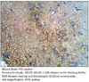
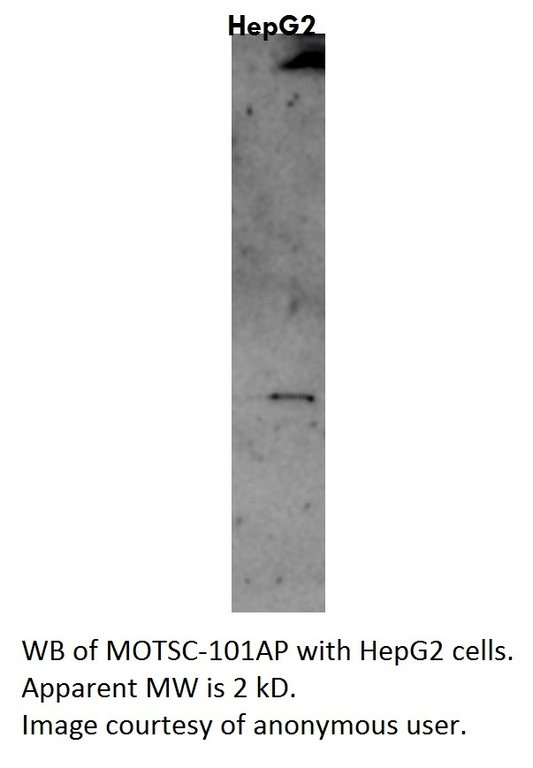
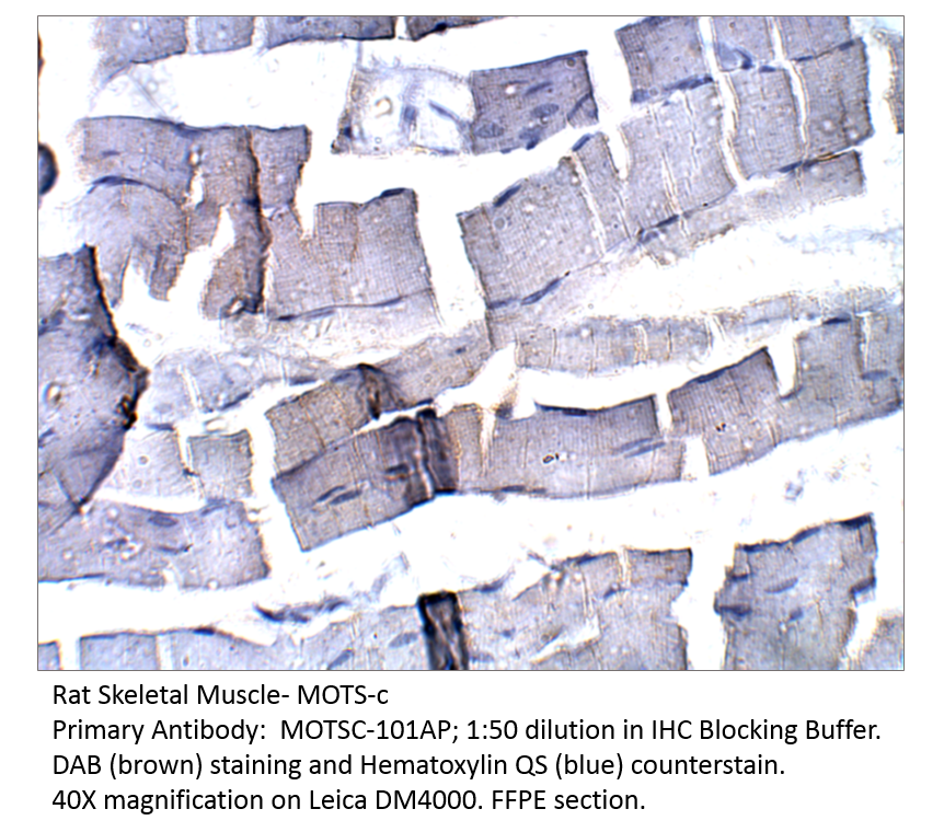
Home page Primary Antibodies Cell Biology Organelles Mitochondria MOTS-c Antibody
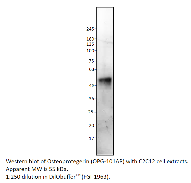
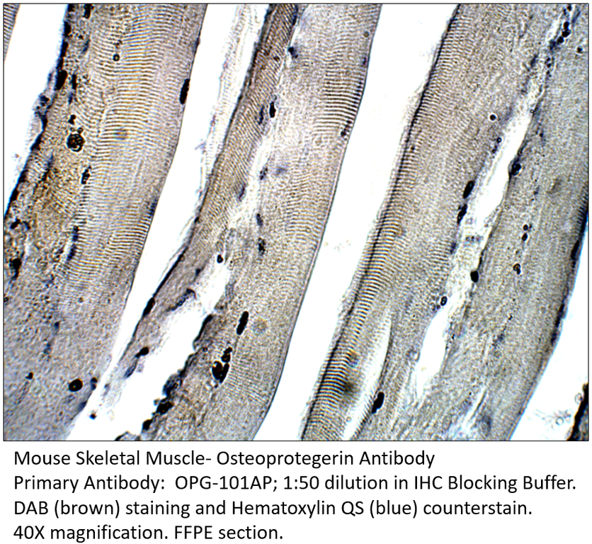
Home page Primary Antibodies Cardiology Osteoprotegerin Antibody
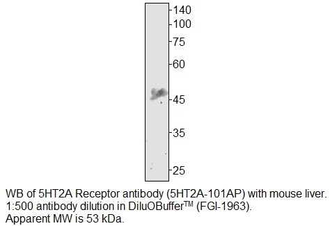
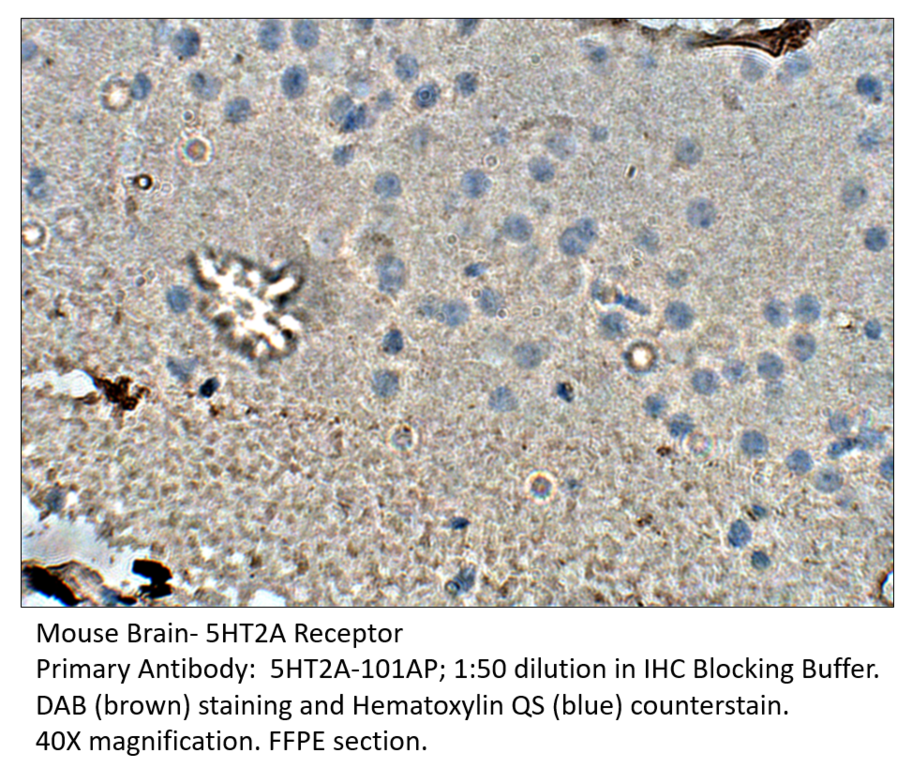
Home page Primary Antibodies Neuroscience Neurotransmission Serotonin Receptors 5HT2A Receptor Antibody
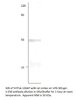
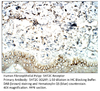
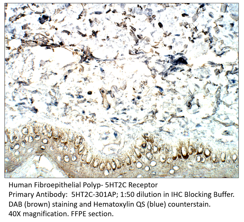
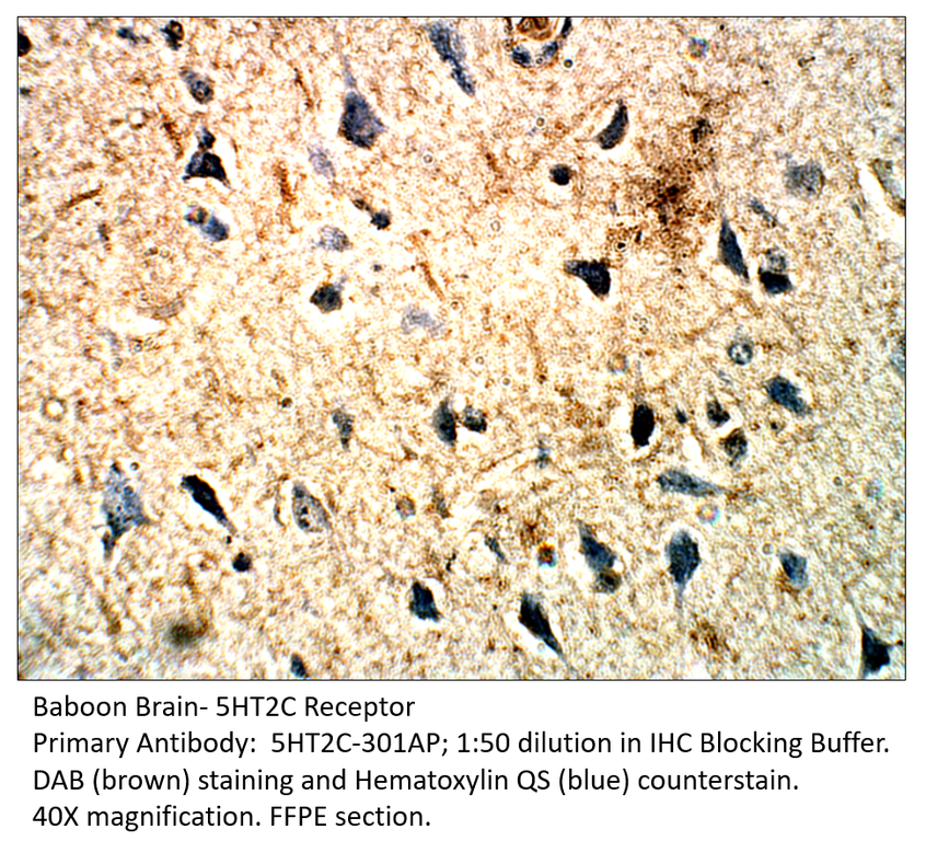
Home page Primary Antibodies Neuroscience Neurotransmission Serotonin Receptors 5HT2C Receptor Antibody
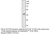
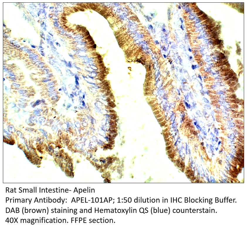
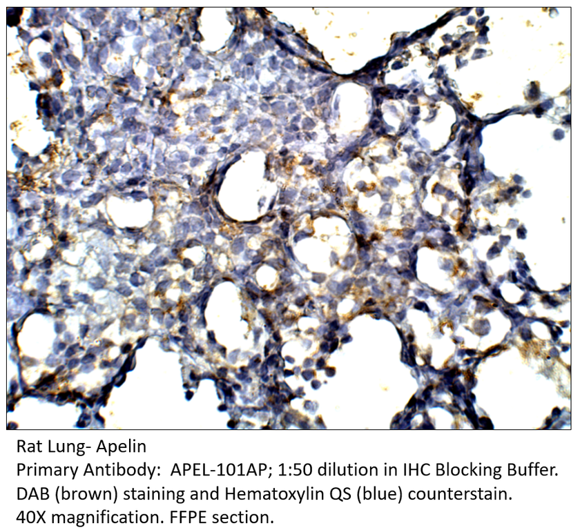
Home page Primary Antibodies Neuroscience Neuropeptides & Hormones Apelin Antibody

Accessories
| Product | Note | Status | Price | |
|---|---|---|---|---|
|
PPKCETA.140-FITC | |||
|
PPKCETA.140-BIOTIN | |||
|
P-PPKCETA.140 | |||
|
PC-PPKCETA | |||
| Display accessory details | ||||
 Phospho-PKC eta Antibody FITC
 Phospho-PKC eta Antibody BIOTIN
 Phospho-PKC eta Blocking Peptide
 Phospho-PKC eta Positive Control
We also recommend
|
PKCETA-101AP
 |
PKCETA-112AP
 |
PKCETA-121AP
 |
|
PPKCETA-240AP
 |
PKCEP-101AP
 |
Browse these categories as well: PKC, Obesity, Primaries

Accessories
| Product | Note | Status | Price | |
|---|---|---|---|---|
|
PPKCETA.240-FITC | |||
|
PPKCETA.240-BIOTIN | |||
|
P-PPKCETA.240 | |||
|
PC-PPKCETA | |||
| Display accessory details | ||||
 Phospho-PKC eta Antibody FITC
 Phospho-PKC eta Antibody BIOTIN
 Phospho-PKC eta Blocking Peptide
We also recommend
|
PPKCETA-140AP
 |
PKCETA-101AP
|
PKCETA-112AP
|
|
PKCETA-121AP
|
PKCEP-101AP
|
Browse these categories as well: PKC, Obesity, Primaries

Accessories
| Product | Note | Status | Price | |
|---|---|---|---|---|
|
PKCETA.112-FITC | |||
|
PKCETA.112-BIOTIN | |||
|
P-PKCETA.112 | |||
|
PC-PKCETA | |||
| Display accessory details | ||||
 PKC eta Antibody FITC
 PKC eta Antibody BIOTIN
 PKC eta Blocking Peptide
 PKC eta Positive Control
We also recommend
|
PPKCETA-140AP
|
PPKCETA-240AP
|
PKCETA-101AP
|
|
PKCETA-121AP
|
PKCEP-101AP
|
Browse these categories as well: PKC, Serine/Threonine Kinases, Obesity, Primaries

Accessories
| Product | Note | Status | Price | |
|---|---|---|---|---|
|
PKCETA.101-FITC | |||
|
PKCETA.101-BIOTIN | |||
|
PC-PKCETA | |||
|
P-PKCETA | |||
| Display accessory details | ||||
 PKC eta Antibody FITC
 PKC eta Antibody BIOTIN
 PKC eta Blocking Peptide
We also recommend
|
PPKCETA-140AP
|
PPKCETA-240AP
|
PKCETA-112AP
|
|
PKCETA-121AP
|
PKCEP-101AP
|
Browse these categories as well: PKC, Serine/Threonine Kinases, Obesity, Primaries

Accessories
| Product | Note | Status | Price | |
|---|---|---|---|---|
|
PKCETA.121-FITC | |||
|
PKCETA.121-BIOTIN | |||
|
P-PKCETA.121 | |||
|
PC-PKCETA | |||
| Display accessory details | ||||
 PKC eta Antibody FITC
 PKC eta Antibody BIOTIN
 PKC eta Blocking Peptide
We also recommend
|
PPKCETA-140AP
|
PKCETA-101AP
|
PKCETA-112AP
|
|
PKCEP-101AP
|
Browse these categories as well: PKC, Serine/Threonine Kinases, Obesity, Primaries

Accessories
| Product | Note | Status | Price | |
|---|---|---|---|---|
|
MOTSC-FITC | |||
|
MOTSC-BIOTIN | |||
|
P-MOTSC | |||
|
PC-MOTSC | |||
| Display accessory details | ||||
 MOTS-c Antibody FITC
 MOTS-c Antibody BIOTIN
 MOTS-c Blocking Peptide
 MOTS-c Positive Control
We also recommend
|
HUMN-101AP
 |
Customers who bought this product also bought
 |
 |
Browse these categories as well: Mitochondria, Obesity, Insulin, Diabetes, Metabolism & Homeostasis, Primaries
Accessories
| Product | Note | Status | Price | |
|---|---|---|---|---|
|
OPG-BIOTIN | |||
|
OPG-FITC | |||
|
P-OPG | |||
|
PC-OPG | |||
| Display accessory details | ||||
 Osteoprotegerin Antibody BIOTIN
 Osteoprotegerin Antibody FITC
 Osteoprotegerin Blocking Peptide
 Osteoprotegerin Positive Control
Browse these categories as well: Cardiology, TNF Superfamily, Cytokines, Bone, Obesity, Primaries
Accessories
| Product | Note | Status | Price | |
|---|---|---|---|---|
|
5HT2A-BIOTIN | |||
|
5HT2A-FITC | |||
|
P-5HT2A | |||
|
PC-5HT2A | |||
| Display accessory details | ||||
 5HT2A Receptor Antibody BIOTIN
 5HT2A Receptor Antibody FITC
 5HT2A Receptor Blocking Peptide
 5HT2A Receptor Positive Control
We also recommend
|
5HT1A-101AP
 |
5HT1B-201AP
 |
5HT2C-301AP
 |
Browse these categories as well: Serotonin Receptors, G-Protein Coupled Receptors, Neurotransmission, Obesity, Primaries
Accessories
| Product | Note | Status | Price | |
|---|---|---|---|---|
|
5HT2C-BIOTIN | |||
|
5HT2C-FITC | |||
|
P-5HT2C | |||
|
PC-5HT2C | |||
| Display accessory details | ||||
 5HT2C Receptor Antibody BIOTIN
 5HT2C Receptor Antibody FITC
 5HT2C Receptor Blocking Peptide
 5HT2C Receptor Positive Control
We also recommend
|
5HT2A-101AP
 |
5HT1A-101AP
|
5HT1B-201AP
|
Browse these categories as well: Serotonin Receptors, G-Protein Coupled Receptors, Neurotransmission, Metabolism & Homeostasis, Obesity, Primaries
Accessories
| Product | Note | Status | Price | |
|---|---|---|---|---|
|
APEL-BIOTIN | |||
|
APEL-FITC | |||
|
P-APEL | |||
|
PC-APEL | |||
| Display accessory details | ||||
 Apelin Antibody BIOTIN
 Apelin Antibody FITC
 Apelin Blocking Peptide
 Apelin Positive Control
We also recommend
|
APLNR-112AP
 |
APLNR-101AP
 |
Browse these categories as well: Neuropeptides & Hormones, G-Protein Coupled Receptors, Cardiology, Obesity, Primaries