Your cart is empty.

Home page Primary Antibodies Signal Transduction Cell Adhesion Occludin Antibody
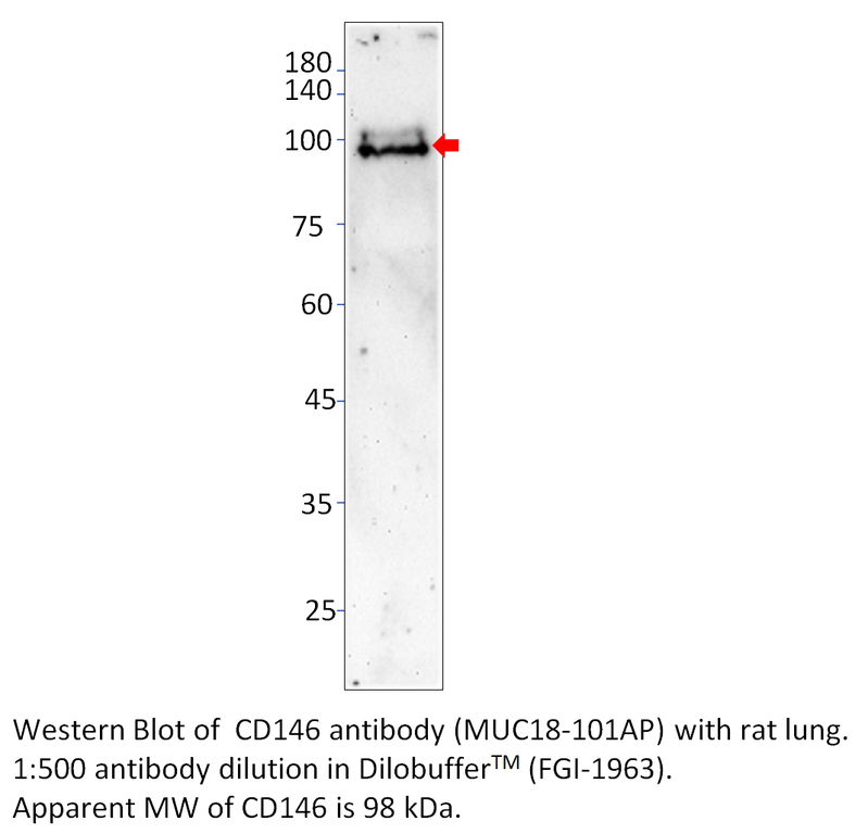
Home page Primary Antibodies Signal Transduction Cell Adhesion CD146 Antibody
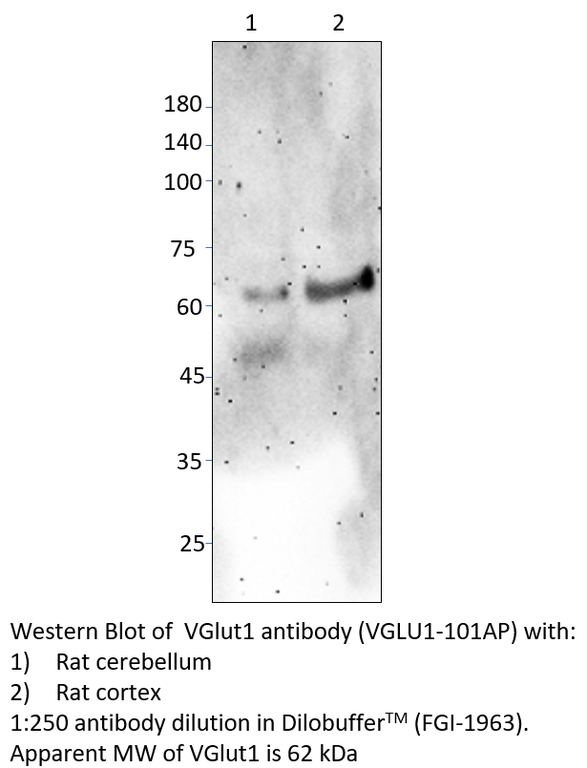
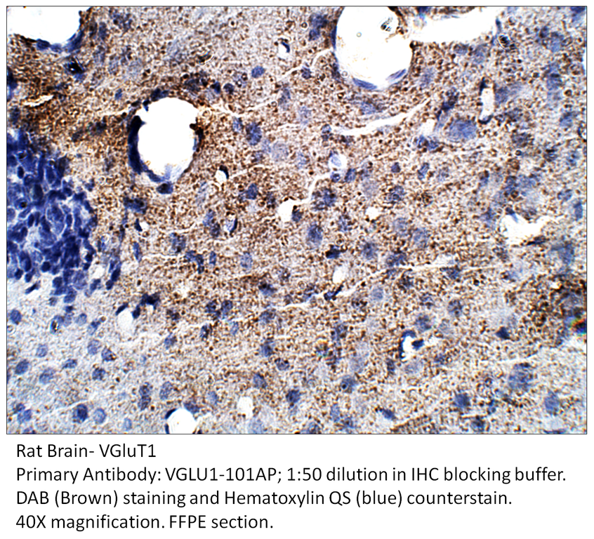
Home page Primary Antibodies Signal Transduction Cell Adhesion VGluT1 Antibody
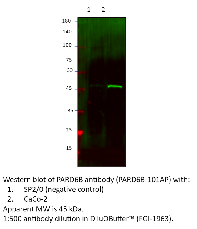
Home page Primary Antibodies Signal Transduction Cell Adhesion PARD6B Antibody
Home page Primary Antibodies Signal Transduction Cell Adhesion PARD6B Antibody
Home page Primary Antibodies Signal Transduction Cytoskeleton / ECM Microfilaments Phospho-Cofilin Antibody

Accessories
| Product | Note | Status | Price | |
|---|---|---|---|---|
|
OCLN-BIOTIN | |||
|
OCLN-FITC | |||
|
P-OCLN | |||
|
PC-OCLN | |||
| Display accessory details | ||||
 Occludin Antibody BIOTIN
 Occludin Antibody FITC
 Occludin Blocking Peptide
 Occludin Positive Control
Browse these categories as well: Cell Adhesion, Cytoskeleton / ECM, Primaries
Accessories
| Product | Note | Status | Price | |
|---|---|---|---|---|
|
MUC18-BIOTIN | |||
|
MUC18-FITC | |||
|
P-MUC18 | |||
|
PC-MUC18 | |||
| Display accessory details | ||||
 Cell Surface Glycoprotein MUC18 Antibody BIOTIN
 Cell Surface Glycoprotein MUC18 Antibody FITC
 Cell Surface Glycoprotein MUC18 Blocking Peptide
 Cell Surface Glycoprotein MUC18 Positive Control
We also recommend
|
MUC-101AP
 |
Browse these categories as well: Cell Adhesion, Cytoskeleton / ECM, Invasion/microenviornment, Stem Cells, Angiogenesis, Primaries
Accessories
| Product | Note | Status | Price | |
|---|---|---|---|---|
|
VGLU1-BIOTIN | |||
|
VGLU1-FITC | |||
|
P-VGLU1 | |||
| Display accessory details | ||||
 VGluT1 Antibody BIOTIN
 VGluT1 Antibody FITC
 VGluT1 Blocking Peptide
Customers who bought this product also bought
 |
 |
 |
 |
Browse these categories as well: Cell Adhesion, Cytoskeleton / ECM, Primaries
Accessories
| Product | Note | Status | Price | |
|---|---|---|---|---|
|
PARD6B-BIOTIN | |||
|
PARD6B-FITC | |||
|
P-PARD6B | |||
|
PC-PARD6B | |||
| Display accessory details | ||||
 PARD6B Antibody BIOTIN
 PARD6B Antibody FITC
 PARD6B Blocking Peptide
 PARD6B Positive Control
We also recommend
|
PARD6B-112AP
 |
Browse these categories as well: Cell Adhesion, Cytoskeleton / ECM, Cell Cycle, Primaries

Accessories
| Product | Note | Status | Price | |
|---|---|---|---|---|
|
PARD6B-112-BIOTIN | |||
|
PARD6B-112-FITC | |||
|
P-PARD6B-112 | |||
|
PC-PARD6B | |||
| Display accessory details | ||||
 PARD6B Antibody BIOTIN
 PARD6B Antibody FITC
 PARD6B Blocking Peptide
We also recommend
|
PARD6B-101AP
 |
Browse these categories as well: Cell Adhesion, Cytoskeleton / ECM, Cell Cycle, Primaries

Accessories
| Product | Note | Status | Price | |
|---|---|---|---|---|
|
PCFL-BIOTIN | |||
|
PCFL-FITC | |||
|
P-PCFL | |||
|
PC-PCFL | |||
| Display accessory details | ||||
 Phospho-Cofilin Antibody BIOTIN
 Phospho-Cofilin Antibody FITC
 Phospho-Cofilin Blocking Peptide
 Phospho-Cofilin Positive Control
We also recommend
|
CFL-101AP
 |
Browse these categories as well: Microfilaments, Growth Factors & Hormones, Cardiology, Cytoskeleton / ECM, Cell Adhesion, Invasion/microenviornment, Actin, Primaries