Your cart is empty.

Home page Primary Antibodies Protein Kinases RIPK2 Antibody
Home page Primary Antibodies Cancer Research Cell Cycle Kinase / Phosphatases RIPK1 Antibody
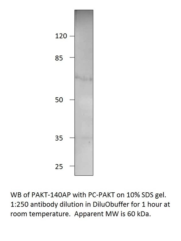
Home page Primary Antibodies Protein Kinases Serine/Threonine Kinases AKT Phospho-AKT1 Antibody
Home page Primary Antibodies Protein Kinases RIPK5 Antibody
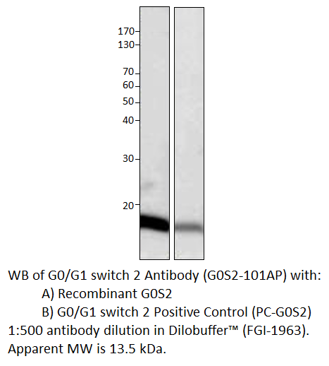
Home page Primary Antibodies Cancer Research Oncoproteins G0/G1 switch 2 Antibody
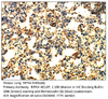
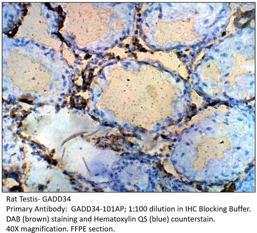
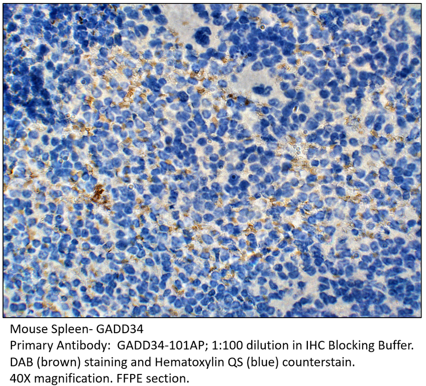
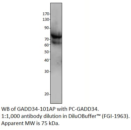
Home page Primary Antibodies Cancer Research Cell Cycle Kinase / Phosphatases GADD34 Antibody
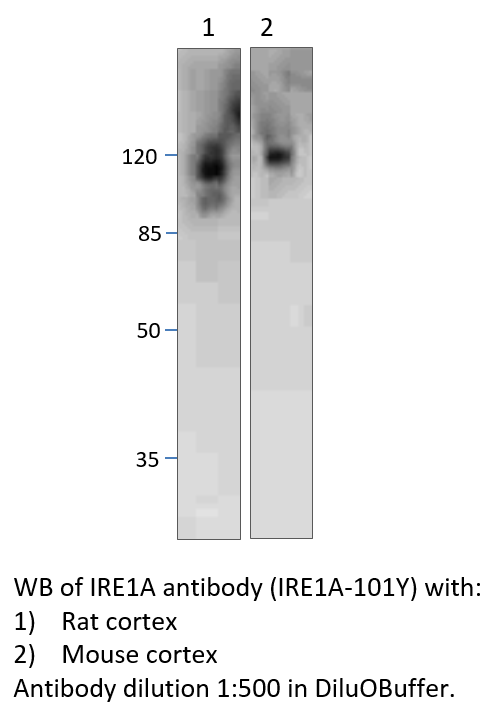
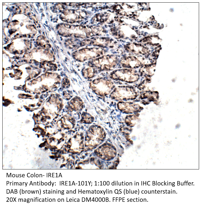
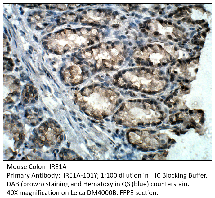
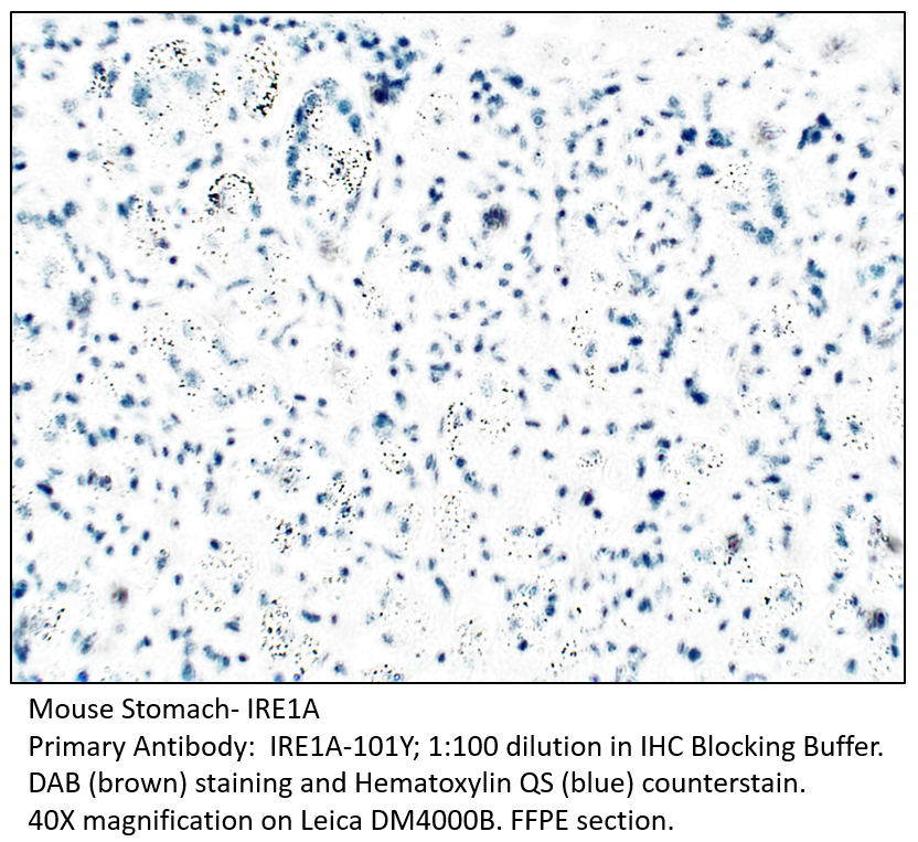
Home page Primary Antibodies Protein Kinases Serine/Threonine Kinases IRE1A Antibody
Home page Primary Antibodies Cell Biology Ubiquitination and Proteinases Ubiquitin E3 Enzymes Hect E3 Ligase ITCH / AIP4 Antibody
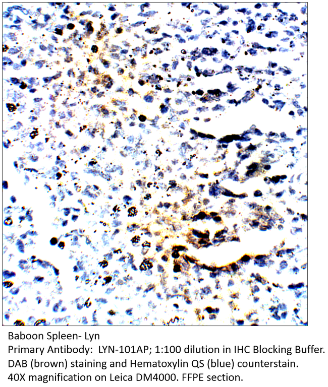
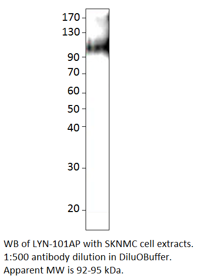
Home page Primary Antibodies Protein Kinases Tyrosine Kinases Src Family Lyn Antibody
Home page Primary Antibodies Protein Kinases Tyrosine Kinases Src Family Phospho-Lyn Antibody

Accessories
| Product | Note | Status | Price | |
|---|---|---|---|---|
|
RIPK2-FITC | |||
|
RIPK2-BIOTIN | |||
|
P-RIPK2 | |||
|
PC-RIPK2 | |||
| Display accessory details | ||||
 RIPK2 Antibody FITC
 RIPK2 Antibody BIOTIN
 RIPK2 Blocking Peptide
 RIPK2 Positive Control
We also recommend
|
RIPK1-101AP
 |
RIPK4-401AP
 |
RIPK5-501AP
 |
Browse these categories as well: Protein Kinases, Serine/Threonine Kinases, Caspases, Receptor Interacting Proteins, Apoptosis, Apoptosis, TLR Signaling, Obesity, Primaries

Accessories
| Product | Note | Status | Price | |
|---|---|---|---|---|
|
RIPK1-FITC | |||
|
RIPK1-BIOTIN | |||
|
P-RIPK1 | |||
|
PC-RIPK1 | |||
| Display accessory details | ||||
 RIPK1 Antibody FITC
 RIPK1 Antibody BIOTIN
 RIPK1 Blocking Peptide
 RIPK1 Positive Control
We also recommend
|
RIPK4-401AP
|
RIPK2-201AP
 |
RIPK5-501AP
|
Browse these categories as well: Kinase / Phosphatases, Apoptosis, Apoptosis, Serine/Threonine Kinases, Receptor Interacting Proteins, Primaries
Accessories
| Product | Note | Status | Price | |
|---|---|---|---|---|
|
PAKT-FITC | |||
|
PAKT-BIOTIN | |||
|
P-PAKT | |||
|
PC-PAKT | |||
| Display accessory details | ||||
 Phospho-AKT1 Antibody FITC
 Phospho-AKT1 Antibody BIOTIN
 Phospho-AKT1 Blocking Peptide
 Phospho-AKT1 Positive Control
Browse these categories as well: AKT, Serine/Threonine Kinases, Cell Cycle, Apoptosis, Nuclear Signaling Pathways, Metabolism, Energy Metabolism, Primaries

Accessories
| Product | Note | Status | Price | |
|---|---|---|---|---|
|
RIPK5-FITC | |||
|
RIPK5-BIOTIN | |||
|
P-RIPK5 | |||
|
PC-RIPK5 | |||
| Display accessory details | ||||
 RIPK5 Antibody FITC
 RIPK5 Antibody BIOTIN
 RIPK5 Blocking Peptide
 RIPK5 Positive Control
We also recommend
|
RIPK4-401AP
|
RIPK1-101AP
|
RIPK2-201AP
|
Browse these categories as well: Protein Kinases, Serine/Threonine Kinases, Receptor Interacting Proteins, Apoptosis, Primaries

Accessories
| Product | Note | Status | Price | |
|---|---|---|---|---|
|
G0S2-FITC | |||
|
G0S2-BIOTIN | |||
|
P-G0S2 | |||
|
PC-G0S2 | |||
| Display accessory details | ||||
 G0/G1 switch 2 Antibody FITC
 G0/G1 switch 2 Antibody BIOTIN
 G0/G1 switch 2 Blocking Peptide
 G0/G1 switch 2 Positive Control
Browse these categories as well: Oncoproteins, Tumor Suppressors, Primaries, Apoptosis, Mitochondria
Accessories
| Product | Note | Status | Price | |
|---|---|---|---|---|
|
GADD34-FITC | |||
|
GADD34-BIOTIN | |||
|
P-GADD34 | |||
|
PC-GADD34 | |||
| Display accessory details | ||||
 GADD34 Antibody FITC
 GADD34 Antibody BIOTIN
 GADD34 Blocking Peptide
 GADD34 Positive Control
We also recommend
|
GADD-101AP
 |
Browse these categories as well: Kinase / Phosphatases, Protein Phosphatases, DNA Damage Response, Primaries, TGF beta, TGF Pathway, Apoptosis
Accessories
| Product | Note | Status | Price | |
|---|---|---|---|---|
|
IRE1A-Y-FITC | |||
|
IRE1A-Y-BIOTIN | |||
|
P-IRE1A-Y | |||
|
PC-IRE1A | |||
| Display accessory details | ||||
 IRE1A Antibody FITC
 IRE1A Antibody BIOTIN
 IRE1A Blocking Peptide
 IRE1A Positive Control
We also recommend
|
Anti-Chicken Secondary
 |
IRE1A-101AP
 |
Browse these categories as well: Serine/Threonine Kinases, Apoptosis, Transcription, Transcription Factors, Protein Kinases, Chicken IgY Primaries, Primaries
Accessories
| Product | Note | Status | Price | |
|---|---|---|---|---|
|
ITCH-FITC | |||
|
ITCH-BIOTIN | |||
|
P-ITCH | |||
|
PC-ITCH | |||
| Display accessory details | ||||
 ITCH / AIP4 Antibody FITC
 ITCH / AIP4 Antibody BIOTIN
 ITCH / AIP4 Blocking Peptide
 ITCH / AIP4 Positive Control
We also recommend
|
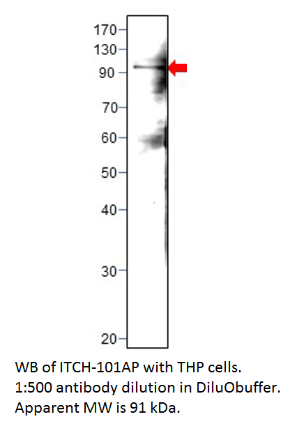
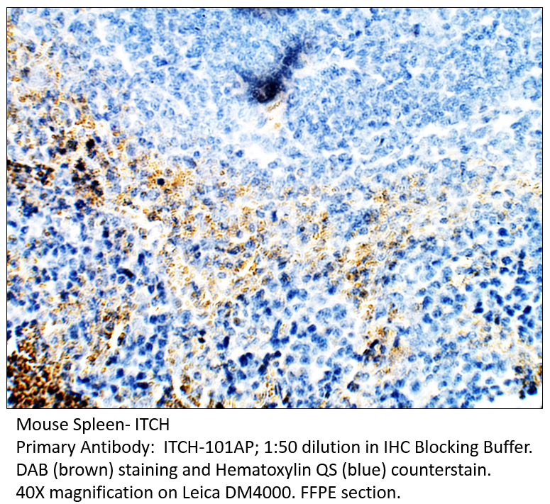
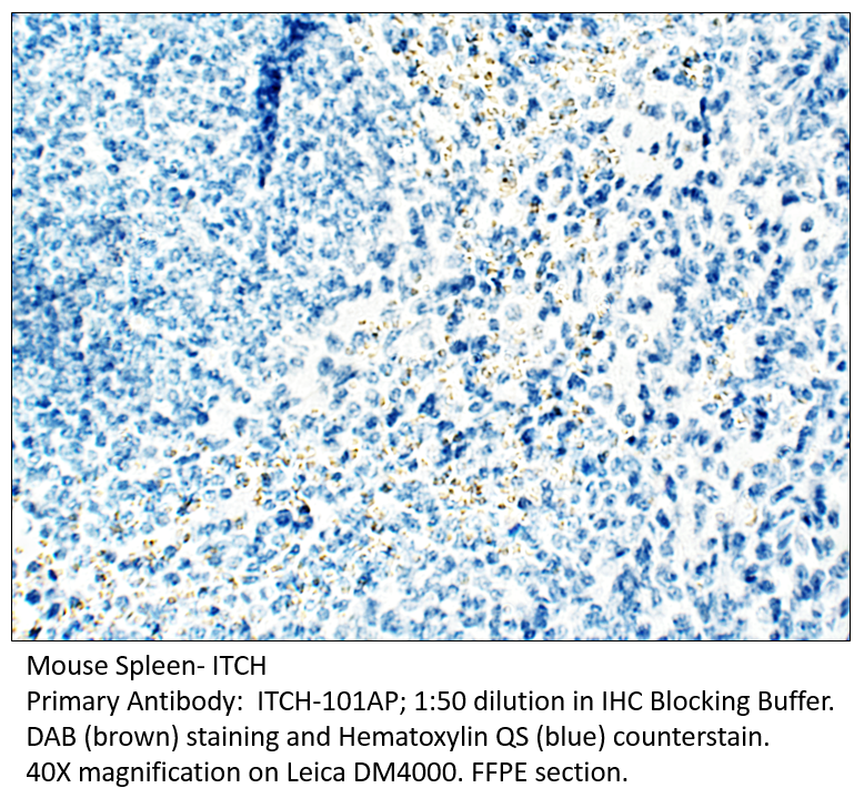
ITCH and TNFAIP3 are part of ubiquitin-editing protein complex ensuring transient nature of inflammatory signaling pathways.
 |
Browse these categories as well: Hect E3 Ligase, Primaries, Transcription, Regulation, Deubiquitination, Apoptosis
Accessories
| Product | Note | Status | Price | |
|---|---|---|---|---|
|
LYN-FITC | |||
|
LYN-BIOTIN | |||
|
P-LYN | |||
|
PC-LYN | |||
| Display accessory details | ||||
 Lyn Antibody FITC
 Lyn Antibody BIOTIN
 Lyn Blocking Peptide
 Lyn Positive Control
We also recommend
|
Lyn acts as the mediator that relays suppressing signals from CXCR4 to beta-2 integrin LFA-1 in hematopoietic precursors.
 |
PLYN-140AP
 |
Browse these categories as well: Src Family, Regulation, Apoptosis, Innate Immunity, Adaptive Immunity, Hematopoietic Progenitors, Growth Factors & Hormones, Growth Factors, DNA Damage Response, Integrins, Protein Kinases, Tyrosine Kinases, Primaries, Receptor Tyrosine Kinases

Accessories
| Product | Note | Status | Price | |
|---|---|---|---|---|
|
PLYN-FITC | |||
|
PLYN-BIOTIN | |||
|
P-PLYN | |||
|
PC-PLYN | |||
| Display accessory details | ||||
 Phospho-Lyn Antibody FITC
 Phospho-Lyn Antibody BIOTIN
 Phospho-Lyn Blocking Peptide
 Phospho-Lyn Positive Control
We also recommend
|
Lyn acts as the mediator that relays suppressing signals from CXCR4 to beta-2 integrin LFA-1 in hematopoietic precursors.
|
LYN-101AP
 |
Browse these categories as well: Src Family, Innate Immunity, Adaptive Immunity, Regulation, Hematopoietic Progenitors, Growth Factors & Hormones, Growth Factors, DNA Damage Response, Integrins, Protein Kinases, Tyrosine Kinases, Apoptosis, Primaries, Receptor Tyrosine Kinases