Your cart is empty.

Home page Primary Antibodies Cell Biology Drosophila melanogaster Smad4 Antibody
Home page Primary Antibodies Cell Biology Drosophila melanogaster Smad4 Antibody
Home page Primary Antibodies Cell Biology Drosophila melanogaster Smad4 Antibody
Home page Primary Antibodies Cell Biology Drosophila melanogaster Smad4 Antibody
Home page Primary Antibodies Metabolism & Homeostasis Hypoxia Visfatin Antibody
Home page Primary Antibodies Metabolism & Homeostasis Hypoxia Visfatin Antibody
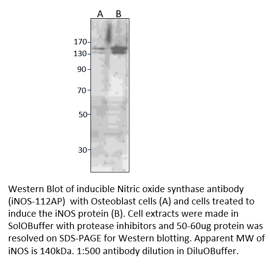
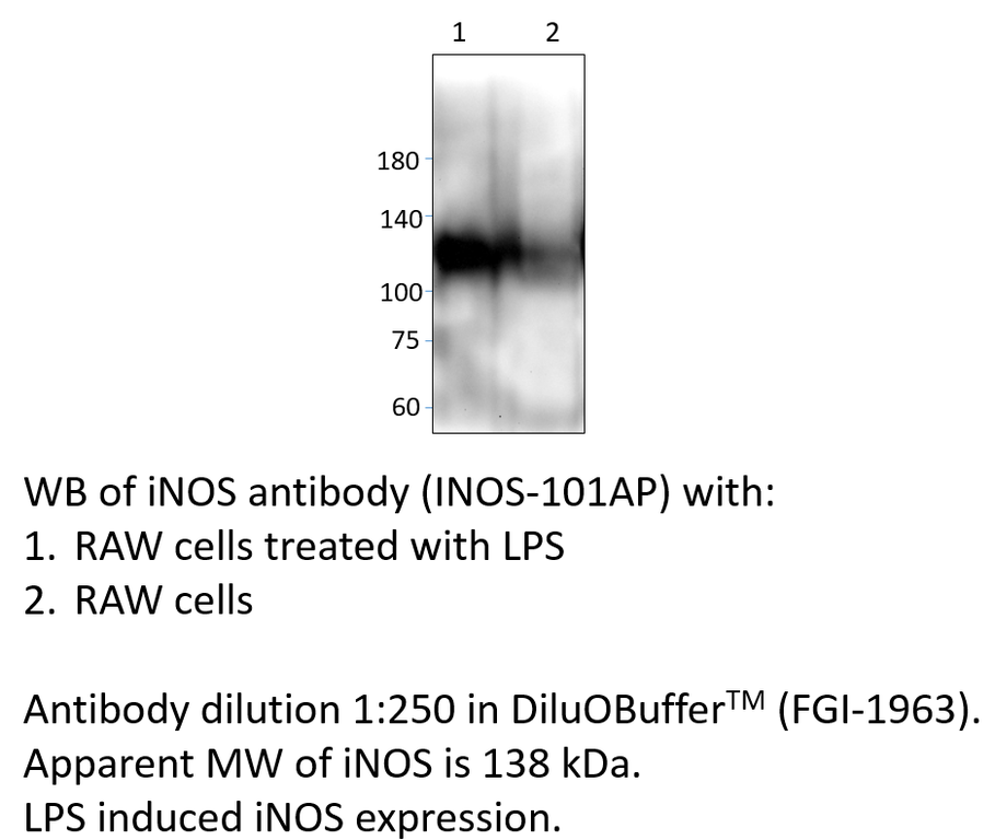
Home page Primary Antibodies Neuroscience Neurotransmission Nitric Oxide iNOS Antibody
Home page Primary Antibodies Neuroscience Neurotransmission Nitric Oxide iNOS Antibody
Home page Primary Antibodies Metabolism & Homeostasis Hypoxia TTC27 Antibody
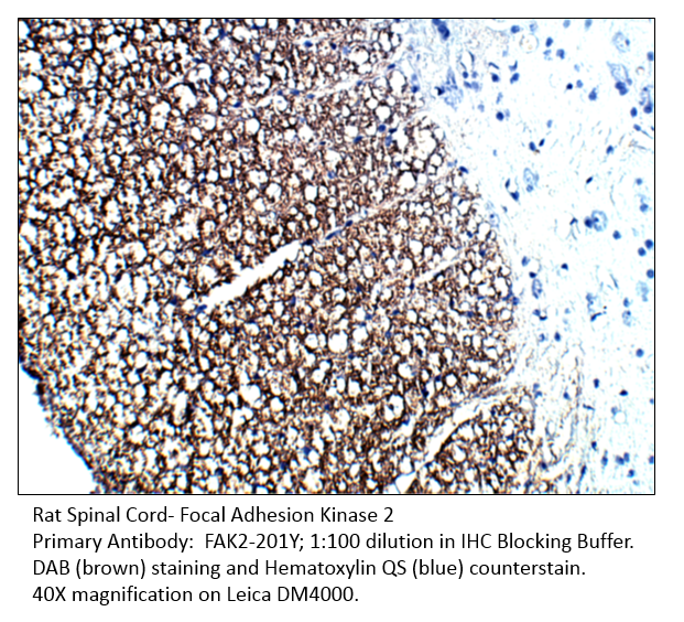
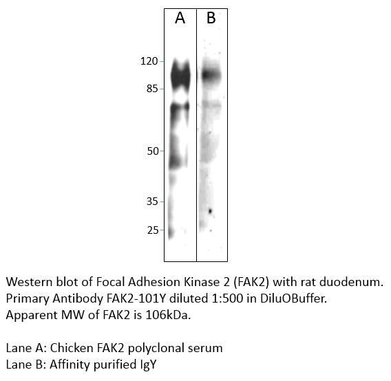
Home page Primary Antibodies Protein Kinases Tyrosine Kinases FAK2 Antibody
Accessories
| Product | Note | Status | Price | |
|---|---|---|---|---|
|
SMAD4.431-FITC | |||
|
SMAD4.431-BIOTIN | |||
|
P-SMAD4.431 | |||
|
PC-SMAD4 | |||
| Display accessory details | ||||
 Smad4 Antibody FITC
 Smad4 Antibody BIOTIN
 Smad4 Blocking Peptide
 Smad4 Antibody Positve Control
We also recommend
|
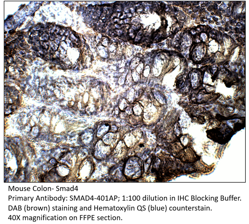
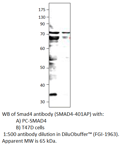
SMAD4-401AP
 |
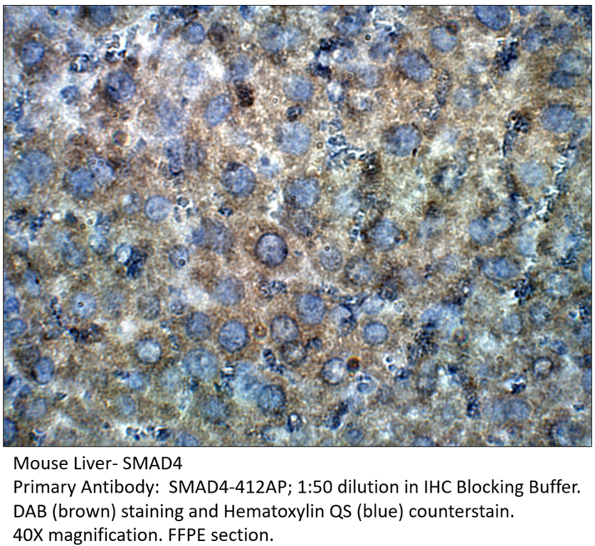
SMAD4-412AP
 |
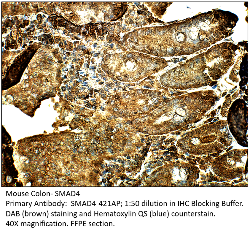
SMAD4-421AP
 |
Browse these categories as well: Drosophila melanogaster, Nuclear Signaling Pathways, TGF beta, TGF Pathway, Tumor Suppressors, Apoptosis, Hypoxia, Cardiology, Metabolism, Metabolism & Homeostasis, Transcription Factors, Transcription, Regulation, Primaries

Accessories
| Product | Note | Status | Price | |
|---|---|---|---|---|
|
SMAD4.412-FITC | |||
|
SMAD4.412-BIOTIN | |||
|
P-SMAD4.412 | |||
|
PC-SMAD4 | |||
| Display accessory details | ||||
 Smad4 Antibody FITC
 Smad4 Antibody BIOTIN
 Smad4 Blocking Peptide
We also recommend
|
SMAD4-401AP
|
SMAD4-421AP
|
SMAD4-431AP
 |
Browse these categories as well: Drosophila melanogaster, Nuclear Signaling Pathways, TGF beta, TGF Pathway, Tumor Suppressors, Apoptosis, Hypoxia, Cardiology, Metabolism, Metabolism & Homeostasis, Transcription Factors, Transcription, Regulation, Primaries

Accessories
| Product | Note | Status | Price | |
|---|---|---|---|---|
|
SMAD4.421-FITC | |||
|
SMAD4.421-BIOTIN | |||
|
P-SMAD4.421 | |||
|
PC-SMAD4 | |||
| Display accessory details | ||||
 Smad4 Antibody FITC
 Smad4 Antibody BIOTIN
 Smad4 Blocking Peptide
We also recommend
|
SMAD4-401AP
|
SMAD4-412AP
|
SMAD4-431AP
|
Browse these categories as well: Drosophila melanogaster, Nuclear Signaling Pathways, TGF beta, TGF Pathway, Tumor Suppressors, Apoptosis, Hypoxia, Cardiology, Metabolism, Metabolism & Homeostasis, Transcription Factors, Transcription, Regulation, Primaries
Accessories
| Product | Note | Status | Price | |
|---|---|---|---|---|
|
SMAD4.401-FITC | |||
|
SMAD4.401-BIOTIN | |||
|
P-SMAD4.401 | |||
|
PC-SMAD4 | |||
| Display accessory details | ||||
 Smad4 Antibody FITC
 Smad4 Antibody BIOTIN
 Smad4 Blocking Peptide
We also recommend
|
SMAD4-412AP
|
SMAD4-421AP
|
SMAD4-431AP
|
Browse these categories as well: Drosophila melanogaster, Nuclear Signaling Pathways, TGF beta, TGF Pathway, Tumor Suppressors, Apoptosis, Hypoxia, Cardiology, Metabolism, Metabolism & Homeostasis, Transcription Factors, Transcription, Regulation, Primaries
Accessories
| Product | Note | Status | Price | |
|---|---|---|---|---|
|
VSFTN-FITC | |||
|
VSFTN-BIOTIN | |||
|
P-VSFTN | |||
|
PC-VSFTN | |||
| Display accessory details | ||||
 Visfatin Antibody FITC
 Visfatin Antibody BIOTIN
 Visfatin Blocking Peptide
 Visfatin Positive Control
We also recommend
|
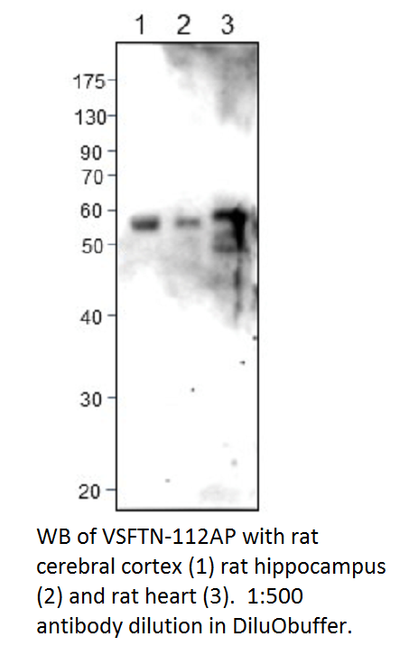
VSFTN-112AP
 |
Browse these categories as well: Hypoxia, Cardiology, B Cells, Cytokines, Metabolism & Homeostasis, Metabolism, Insulin, Primaries

Accessories
| Product | Note | Status | Price | |
|---|---|---|---|---|
|
VSFTN.c-FITC | |||
|
VSFTN.c-BIOTIN | |||
|
P-VSFTN.c | |||
|
PC-VSFTN | |||
| Display accessory details | ||||
 Visfatin Antibody FITC
 Visfatin Antibody BIOTIN
 Visfatin Blocking Peptide
We also recommend
|
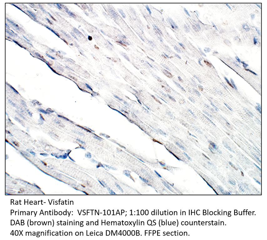
VSFTN-101AP
 |
Browse these categories as well: Hypoxia, Cardiology, B Cells, Cytokines, Metabolism, Metabolism & Homeostasis, Insulin, Primaries
Accessories
| Product | Note | Status | Price | |
|---|---|---|---|---|
|
iNOS.112-BIOTIN | |||
|
iNOS.112-FITC | |||
|
P-iNOS.112 | |||
|
PC-iNOS | |||
| Display accessory details | ||||
 iNOS Antibody BIOTIN
 iNOS Antibody FITC
 iNOS Blocking Peptide
 iNOS Positive Control
We also recommend
|
iNOS1-101AP
 |
Browse these categories as well: Nitric Oxide, Hypoxia, Metabolism, Primaries
Accessories
| Product | Note | Status | Price | |
|---|---|---|---|---|
|
iNOS-BIOTIN | |||
|
iNOS-FITC | |||
|
P-iNOS | |||
|
PC-iNOS | |||
| Display accessory details | ||||
 iNOS Antibody BIOTIN
 iNOS Antibody FITC
 iNOS Blocking Peptide
We also recommend
|
iNOS2-112AP
 |
Browse these categories as well: Nitric Oxide, Hypoxia, Metabolism, Primaries

Accessories
| Product | Note | Status | Price | |
|---|---|---|---|---|
|
TTC27.112-FITC | |||
|
TTC27.112-BIOTIN | |||
|
P-TTC27.112 | |||
|
PC-TTC27 | |||
| Display accessory details | ||||
 TTC27 Antibody FITC
 TTC27 Antibody BIOTIN
 TTC27 Blocking Peptide
 TTC27 Positive Control
We also recommend
|
TTC27-101AP
 |
Browse these categories as well: Hypoxia, Cardiology, Metabolism & Homeostasis, Primaries
Accessories
| Product | Note | Status | Price | |
|---|---|---|---|---|
|
FAK2-Y-FITC | |||
|
FAK2-Y-BIOTIN | |||
|
P-FAK2-Y | |||
|
PC-FAK2 | |||
| Display accessory details | ||||
 FAK2 Antibody FITC
 FAK2 Antibody BIOTIN
 FAK2 Blocking Peptide
 FAK2 Positive Control
We also recommend
|
Anti-Chicken Secondary
 |
FAK1-101Y
 |
Browse these categories as well: Tyrosine Kinases, Hypoxia, Metabolism, Primaries, Chicken IgY Primaries, Non-Receptor Tyrosine Kinases