Your cart is empty.

Home page Primary Antibodies Cancer Research Invasion/microenviornment Apoptosis Lamin B1 antibody - Nuclear Envelope Marker Antibody
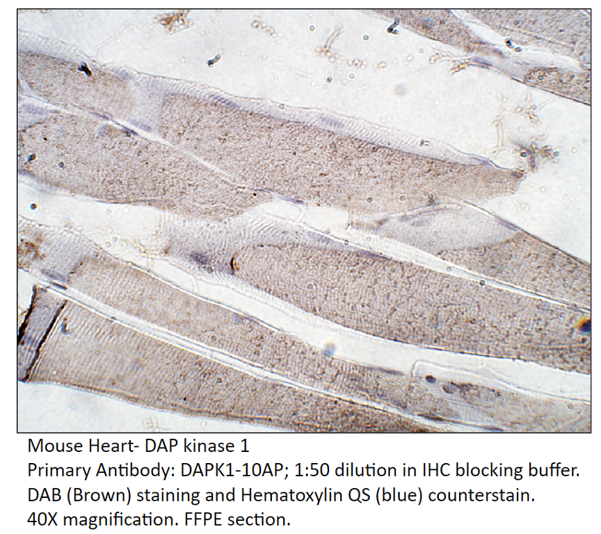
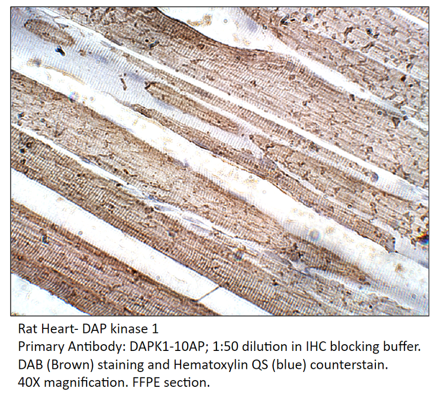
Home page Primary Antibodies Cancer Research Invasion/microenviornment Apoptosis Death receptors & ligands DAP Kinase 1 Antibody
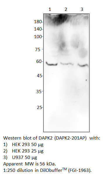
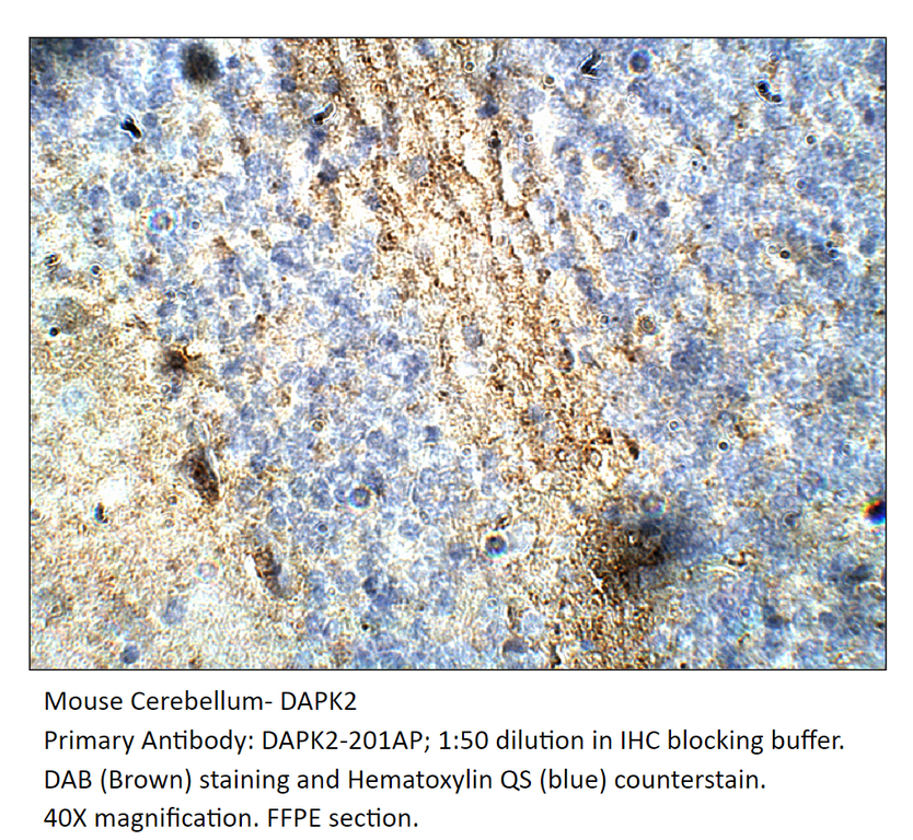
Home page Primary Antibodies Protein Kinases DAPK2 Antibody
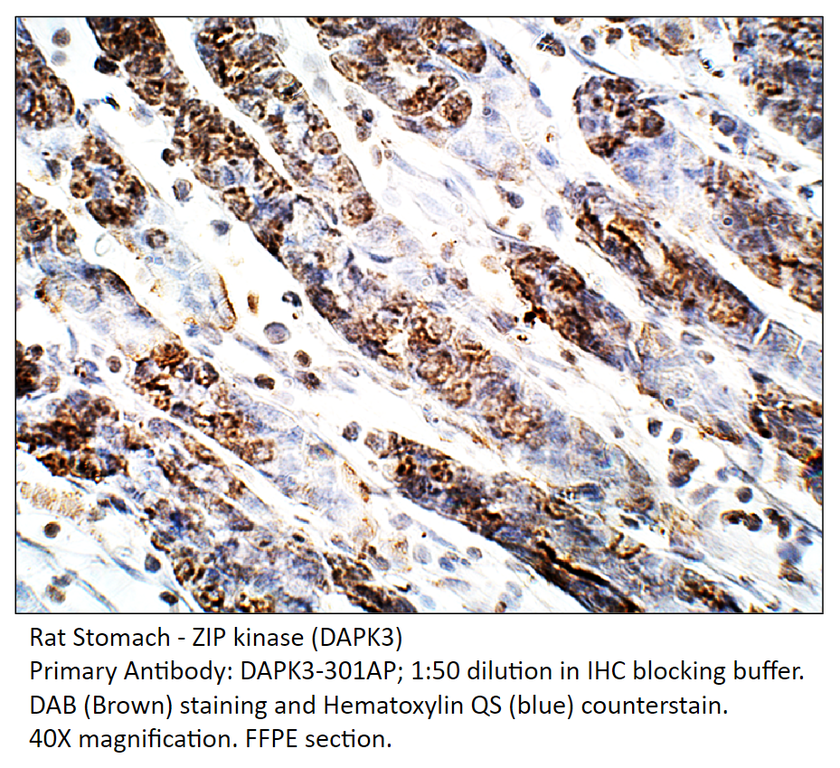
Home page Primary Antibodies Protein Kinases ZIP Kinase Antibody
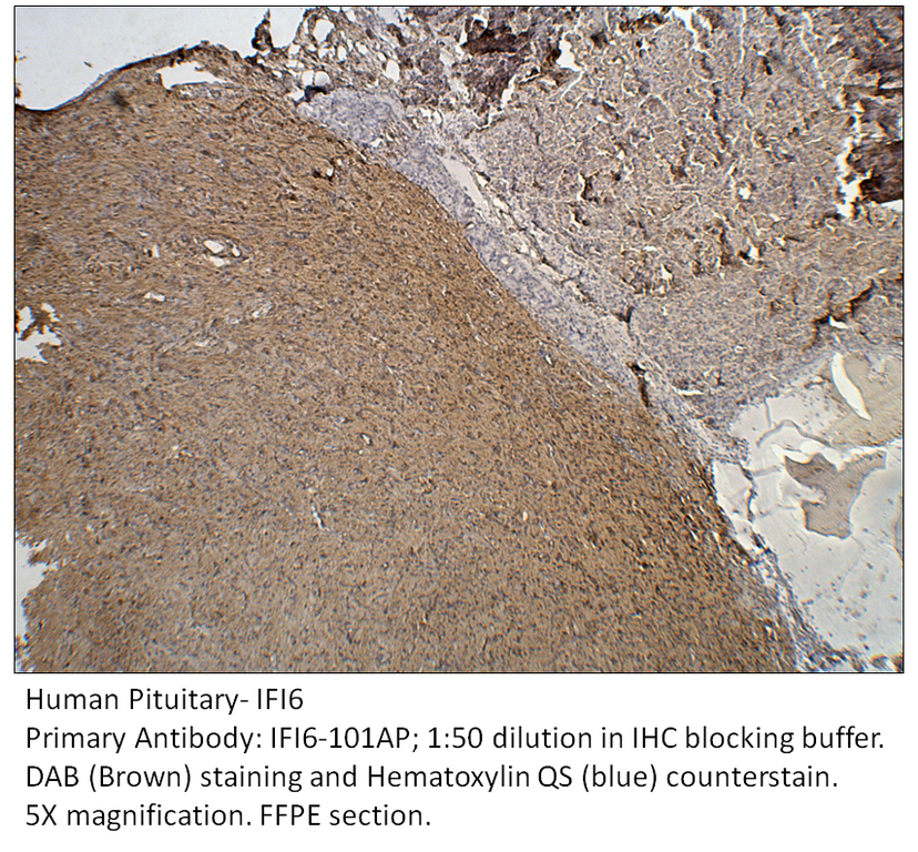
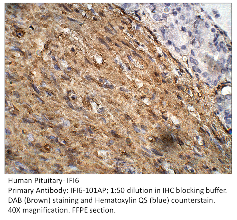
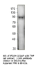
Home page Primary Antibodies Cell Biology Organelles Mitochondria IFI6 Antibody
Home page Primary Antibodies Protein Kinases Serine/Threonine Kinases MLKL Antibody
Home page Primary Antibodies Protein Kinases Serine/Threonine Kinases Phospho-MLKL Antibody
Home page Primary Antibodies Neuroscience Neurodegenerative Disease Alzheimer's Research Amyloid Humanin Antibody
Home page Primary Antibodies Neuroscience Neurodegenerative Disease Alzheimer's Research Amyloid Humanin Antibody
Lamin B1 antibody - Nuclear Envelope Marker Antibody
Product no.: LMNB1-101AP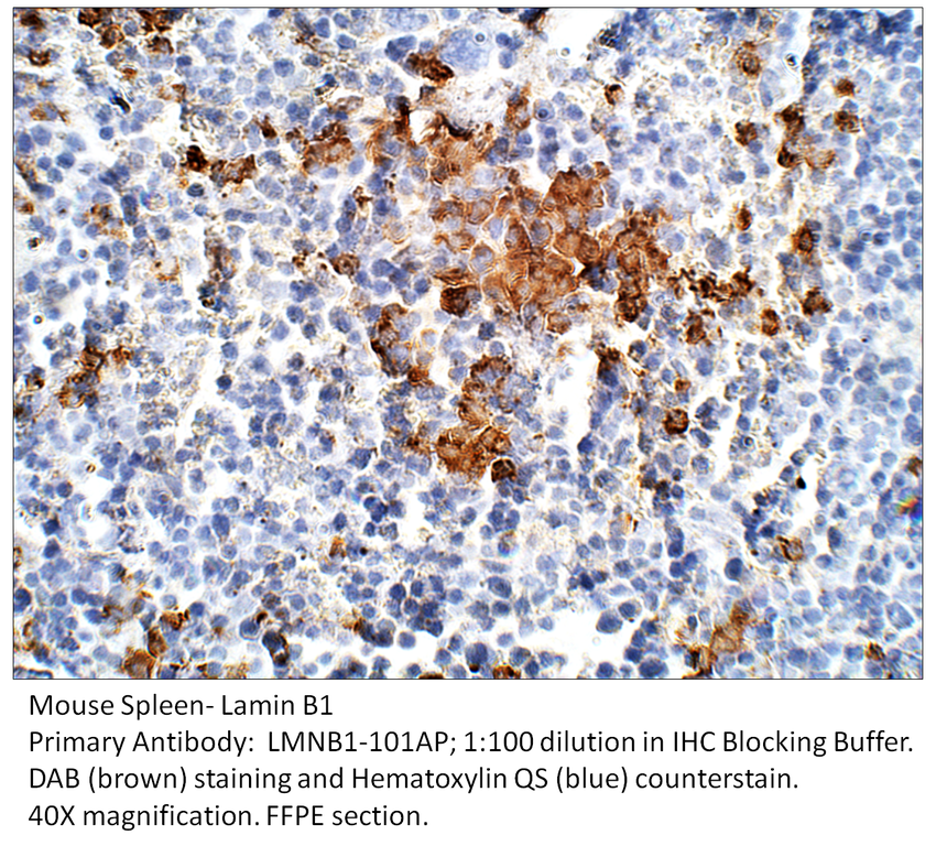
Accessories
| Product | Note | Status | Price | |
|---|---|---|---|---|
|
LMNB1-BIOTIN | |||
|
LMNB1-FITC | |||
|
P-LMNB1 | |||
|
PC-LMNB1 | |||
| Display accessory details | ||||
 Lamin B1 antibody - Nuclear Envelope Marker Antibody BIOTIN
 Lamin B1 antibody - Nuclear Envelope Marker Antibody FITC
 Lamin B1 antibody - Nuclear Envelope Marker Blocking Peptide
 Lamin B1 antibody - Nuclear Envelope Marker Positive Control
Browse these categories as well: Apoptosis, Apoptosis, Cell markers & CAMs, Tags and Cell Markers, Intermediate Filaments, Death receptors & ligands, Primaries
Accessories
| Product | Note | Status | Price | |
|---|---|---|---|---|
|
DAPK1-BIOTIN | |||
|
PC-DAPK1 | |||
|
P-DAPK1 | |||
|
DAPK1-FITC | |||
| Display accessory details | ||||
 DAP Kinase 1 Antibody BIOTIN
 DAP Kinase 1 Positive Control
 DAP Kinase 1 Blocking Peptide
 DAP Kinase 1 Antibody FITC
We also recommend
|
DAPK2-201AP
 |
DAPK1-101AP
 |
Browse these categories as well: Death receptors & ligands, Apoptosis, Protein Kinases, Calcium Binding, Signal Transduction, Signal Transduction, Oncoproteins, Primaries
Accessories
| Product | Note | Status | Price | |
|---|---|---|---|---|
|
DAPK2-BIOTIN | |||
|
PC-DAPK2 | |||
|
P-DAPK2 | |||
|
DAPK2-FITC | |||
| Display accessory details | ||||
 DAPK2 Antibody BIOTIN
 DAPK2 Positive Control
 DAPK2 Blocking Peptide
 DAPK2 Antibody FITC
We also recommend
|
DAPK2-201AP
 |
DAPK1-101AP
|
Browse these categories as well: Protein Kinases, Apoptosis, Primaries
Accessories
| Product | Note | Status | Price | |
|---|---|---|---|---|
|
DAPK3-BIOTIN | |||
|
PC-DAPK3 | |||
|
P-DAPK3 | |||
|
DAPK3-FITC | |||
| Display accessory details | ||||
 ZIP Kinase Antibody BIOTIN
 ZIP Kinase Positive Control
 ZIP Kinase Blocking Peptide
 ZIP Kinase Antibody FITC
We also recommend
|
DAPK2-201AP
|
DAPK1-101AP
|
Browse these categories as well: Protein Kinases, Apoptosis, Apoptosis, Signal Transduction, Signal Transduction, Primaries
Accessories
| Product | Note | Status | Price | |
|---|---|---|---|---|
|
IFI6-BIOTIN | |||
|
PC-IFI6 | |||
|
P-IFI6 | |||
|
IFI6-FITC | |||
| Display accessory details | ||||
 IFI6 Antibody BIOTIN
 IFI6 Positive Control
 IFI6 Blocking Peptide
 IFI6 Antibody FITC
We also recommend
|
IFI27-101AP
 |
IFI16H-101AP
 |
IFI27-112AP
 |
Browse these categories as well: Mitochondria, Metabolism & Homeostasis, Apoptosis, Apoptosis, Primaries

Accessories
| Product | Note | Status | Price | |
|---|---|---|---|---|
|
MLKL-BIOTIN | |||
|
MLKL-FITC | |||
|
P-MLKL | |||
|
PC-MLKL | |||
| Display accessory details | ||||
 MLKL Antibody BIOTIN
 MLKL Antibody FITC
 MLKL Blocking Peptide
 MLKL Positive Control
We also recommend
|
PMLKL-140AP
 |
Browse these categories as well: Serine/Threonine Kinases, Apoptosis, Apoptosis, Primaries

Accessories
| Product | Note | Status | Price | |
|---|---|---|---|---|
|
PMLKL-BIOTIN | |||
|
PMLKL-FITC | |||
|
P-PMLKL | |||
|
PC-PMLKL | |||
| Display accessory details | ||||
 Phospho-MLKL Antibody BIOTIN
 Phospho-MLKL Antibody FITC
 Phospho-MLKL Blocking Peptide
 Phospho-MLKL Positive Control
We also recommend
|
MLKL-101AP
 |
Browse these categories as well: Serine/Threonine Kinases, Apoptosis, Apoptosis, Primaries

Accessories
| Product | Note | Status | Price | |
|---|---|---|---|---|
|
HUMN-112Y-BIOTIN | |||
|
HUMN-112Y-FITC | |||
|
P-HUMN-112Y | |||
|
PC-HUMN | |||
| Display accessory details | ||||
 Humanin Antibody BIOTIN
 Humanin Antibody FITC
 Humanin Blocking Peptide
 Humanin Positive Control
We also recommend
|
HUMN-101AP
 |
HUMN-112AP
 |
Browse these categories as well: Amyloid, Apoptosis, Chicken IgY Primaries, Primaries

Accessories
| Product | Note | Status | Price | |
|---|---|---|---|---|
|
HUMN-112-BIOTIN | |||
|
HUMN-112-FITC | |||
|
P-HUMN-112 | |||
|
PC-HUMN | |||
| Display accessory details | ||||
 Humanin Antibody BIOTIN
 Humanin Antibody FITC
 Humanin Blocking Peptide
We also recommend
|
HUMN-101AP
|
HUMN-112Y
 |
Customers who bought this product also bought
 |
Browse these categories as well: Amyloid, Apoptosis, Primaries