Your cart is empty.

Home page Primary Antibodies Cell Biology C11orf1 Antibody
Home page Primary Antibodies Cell Biology C11orf1 Antibody
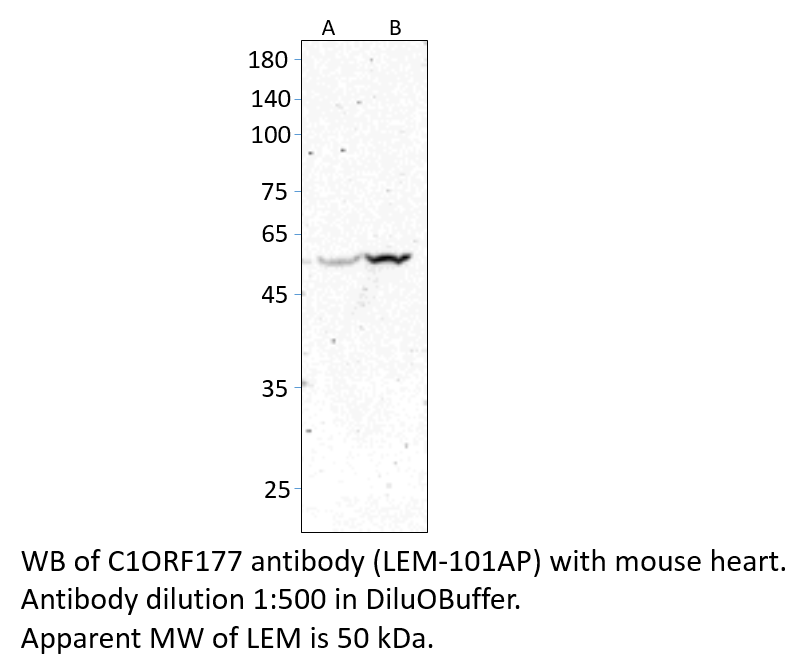
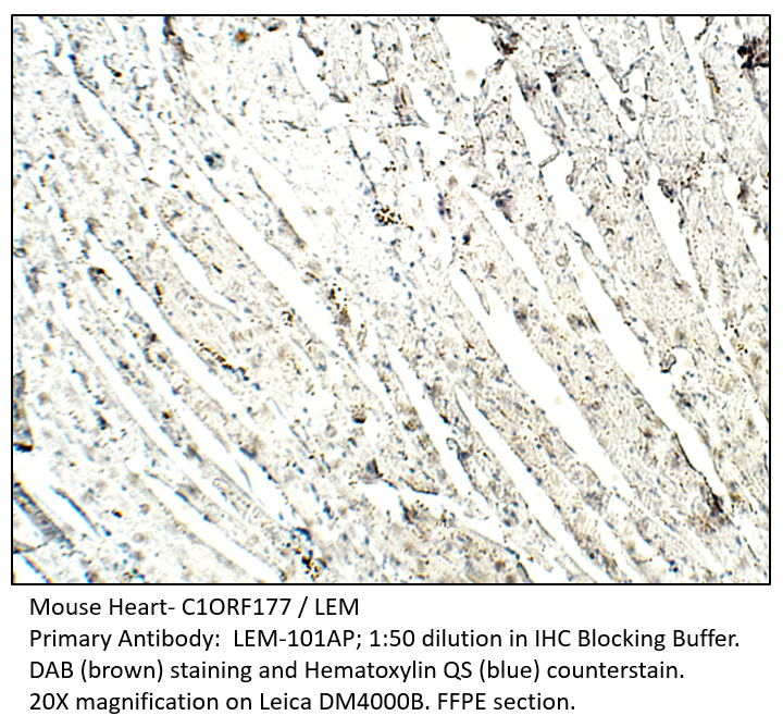
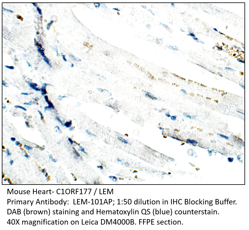
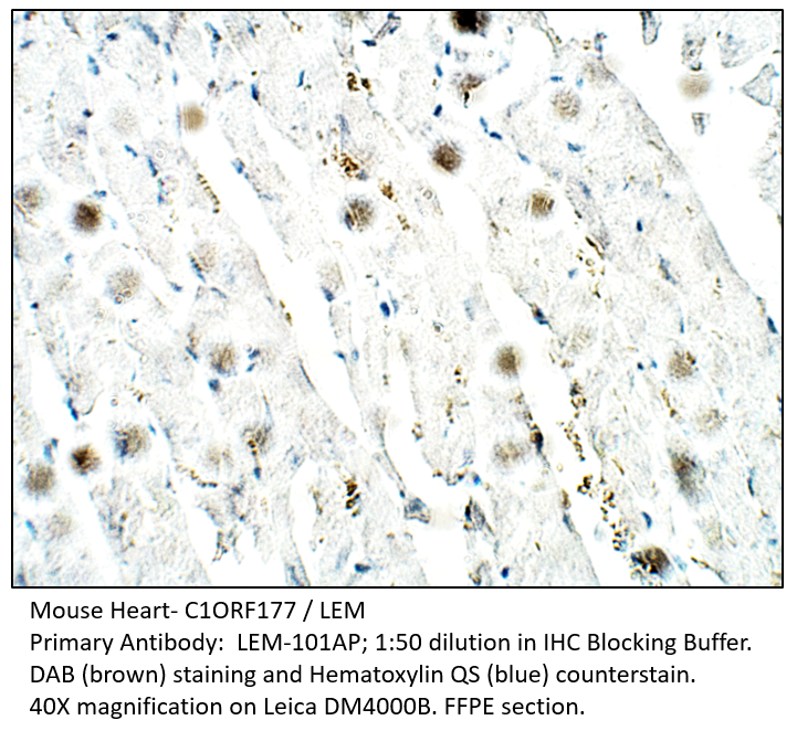
Home page Primary Antibodies Cell Biology C1ORF177 Antibody
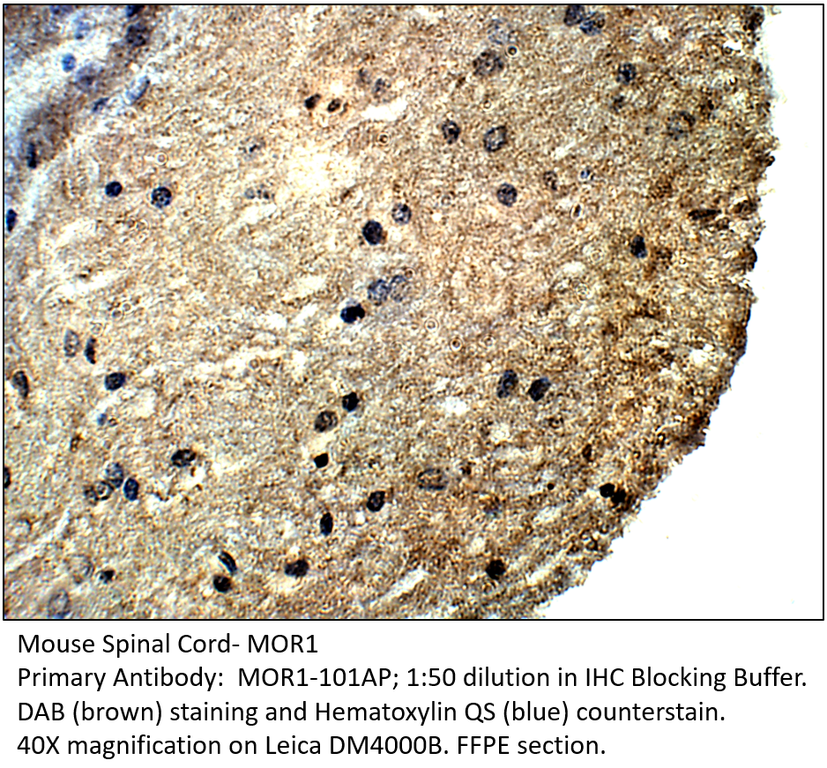
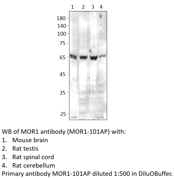
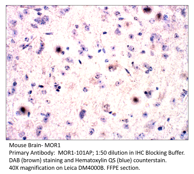
Home page Primary Antibodies Neuroscience MOR1 Antibody
Home page Primary Antibodies Microbiology Virus Syncytin A Antibody
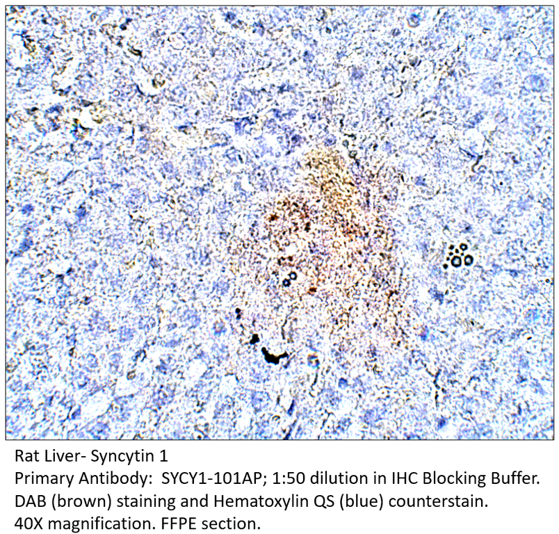
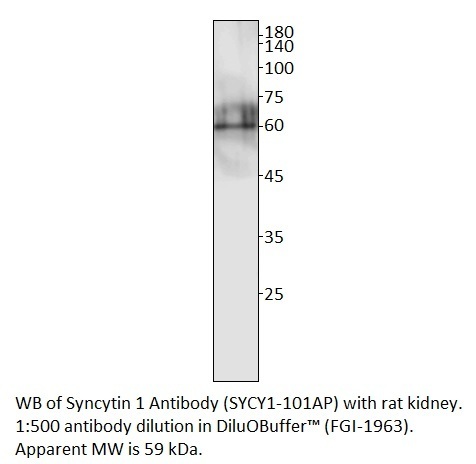
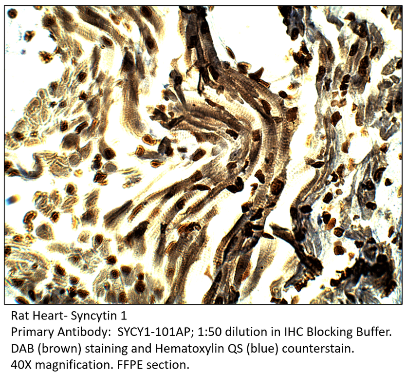
Home page Primary Antibodies Microbiology Virus Syncytin 1 Antibody
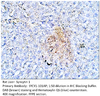
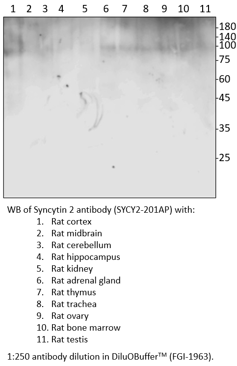
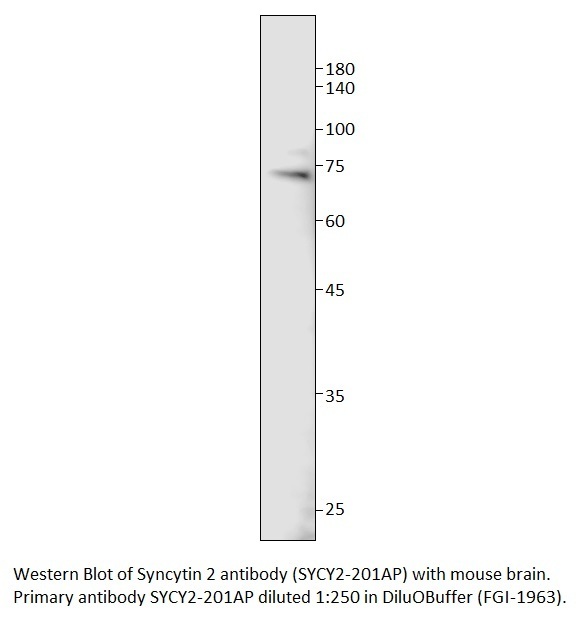
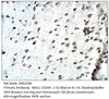
Home page Primary Antibodies Microbiology Virus Syncytin 2 Antibody
Home page Primary Antibodies Microbiology Virus Syncytin 2 Antibody
Home page Primary Antibodies Neuroscience Neurotransmission Dopamine Receptors NSG2 Antibody
Home page Primary Antibodies Neuroscience Sensory Systems Auditory TMC2 Antibody

Accessories
| Product | Note | Status | Price | |
|---|---|---|---|---|
|
FLJ.m-FITC | |||
|
FLJ.m-BIOTIN | |||
|
P-FLJ.m | |||
| Display accessory details | ||||
 C11orf1 Antibody FITC FLJ-112AP
 C11orf1 Antibody BIOTIN FLJ-112AP
 C11orf1 Blocking Peptide FLJ-112AP
We also recommend
|
FLJ-101AP
 |
FLJ-121AP
 |
Browse these categories as well: Cell Biology, Diagnostic Markers, Primaries

Accessories
| Product | Note | Status | Price | |
|---|---|---|---|---|
|
FLJ.c-FITC | |||
|
FLJ.c-BIOTIN | |||
|
P-FLJ.c | |||
| Display accessory details | ||||
 C11orf1 Antibody FITC FLJ-121AP
 C11orf1 Antibody BIOTIN FLJ-121AP
 C11orf1 Blocking Peptide FLJ-121AP
We also recommend
|
FLJ-112AP
 |
FLJ-101AP
|
Browse these categories as well: Cell Biology, Diagnostic Markers, Primaries
Accessories
| Product | Note | Status | Price | |
|---|---|---|---|---|
|
LEM-FITC | |||
|
LEM-BIOTIN | |||
|
P-LEM | |||
|
PC-LEM | |||
| Display accessory details | ||||
 C1ORF177 Antibody FITC
 C1ORF177 Antibody BIOTIN
 C1ORF177 Blocking Peptide
 C1ORF177 Positive Control
Browse these categories as well: Cell Biology, Primaries
Accessories
| Product | Note | Status | Price | |
|---|---|---|---|---|
|
MOR1-BIOTIN | |||
|
MOR1-FITC | |||
|
P-MOR1 | |||
|
PC-MOR1 | |||
| Display accessory details | ||||
 MOR1 Antibody BIOTIN
 MOR1 Antibody FITC
 MOR1 Blocking Peptide
 MOR1 Positive Control
Browse these categories as well: Neuroscience, Neurotransmission, Sensory Systems, Cell Biology, Primaries

Accessories
| Product | Note | Status | Price | |
|---|---|---|---|---|
|
SYCY1-BIOTIN | |||
|
SYCY1-FITC | |||
|
P-SYCY1 | |||
|
PC-SYCY1 | |||
| Display accessory details | ||||
 Syncytin A Antibody BIOTIN
 Syncytin A Antibody FITC
 Syncytin A Blocking Peptide
 Syncytin A Positive Control
We also recommend
|
SYCY1-112AP
 |
SYCY2-201AP
 |
SYCY2-212AP
 |
Browse these categories as well: Virus, Cell Biology, Primaries

Accessories
| Product | Note | Status | Price | |
|---|---|---|---|---|
|
SYCY1-112-BIOTIN | |||
|
SYC1-112-FITC | |||
|
P-SYCY1-112 | |||
|
PC-SYCY1 | |||
| Display accessory details | ||||
 Syncytin 1 Antibody BIOTIN
 Syncytin 1 Antibody FITC
 Syncytin 1 Blocking Peptide
We also recommend
|
SYCY1-101AP
 |
SYCY2-201AP
|
SYCY2-212AP
|
Browse these categories as well: Virus, Cell Biology, Primaries
Accessories
| Product | Note | Status | Price | |
|---|---|---|---|---|
|
SYCY2-BIOTIN | |||
|
SYCY2-FITC | |||
|
P-SYCY2 | |||
|
PC-SYCY2 | |||
| Display accessory details | ||||
 Syncytin 2 Antibody BIOTIN
 Syncytin 2 Antibody FITC
 Syncytin 2 Blocking Peptide
 Syncytin 2 Positive Control
We also recommend
|
SYCY1-101AP
|
SYCY1-112AP
|
SYCY2-212AP
|
Browse these categories as well: Virus, Cell Biology, Primaries

Accessories
| Product | Note | Status | Price | |
|---|---|---|---|---|
|
SYCY2-212-BIOTIN | |||
|
SYCY2-212-FITC | |||
|
P-SYCY2-212 | |||
|
PC-SYCY2 | |||
| Display accessory details | ||||
 Syncytin 2 Antibody BIOTIN
 Syncytin 2 Antibody FITC
 Syncytin 2 Blocking Peptide
We also recommend
|
SYCY1-101AP
|
SYCY1-112AP
|
SYCY2-201AP
|
Browse these categories as well: Virus, Cell Biology, Primaries


Accessories
| Product | Note | Status | Price | |
|---|---|---|---|---|
|
NSG2-BIOTIN | |||
|
NSG2-FITC | |||
|
P-NSG2 | |||
|
PC-NSG2 | |||
| Display accessory details | ||||
 NSG2 Antibody BIOTIN
 NSG2 Antibody FITC
 NSG2 Blocking Peptide
 NSG2 Positive Control
We also recommend
|
NSG1-101AP
 |
Browse these categories as well: Dopamine Receptors, G-Protein Coupled Receptors, Cell Biology, Primaries

Accessories
| Product | Note | Status | Price | |
|---|---|---|---|---|
|
TMC2-BIOTIN | |||
|
PC-TMC2 | |||
|
P-TMC2 | |||
|
TMC2-FITC | |||
| Display accessory details | ||||
 TMC2 Antibody BIOTIN
 TMC2 Positive Control
 TMC2 Blocking Peptide
 TMC2 Antibody FITC
We also recommend
|
TMC1-101AP
 |
Browse these categories as well: Auditory, Cell Biology, Primaries